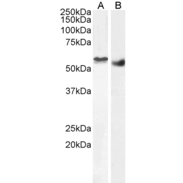
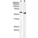
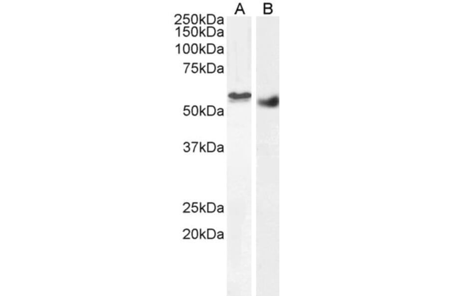
STK35 expression in nuclear HEK293 (A) and Human Breast cancer (B) lysates analyzed by western blot. Cells were lysed in RIPA buffer and 35µg protein was run per lane. Primary incubation was performed with Anti-STK35 Antibody (A84004) at 2µg/ml (A) or 1µg/ml (B) and detected by chemiluminescence.
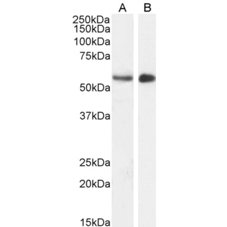
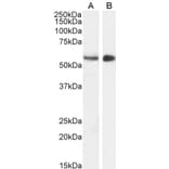
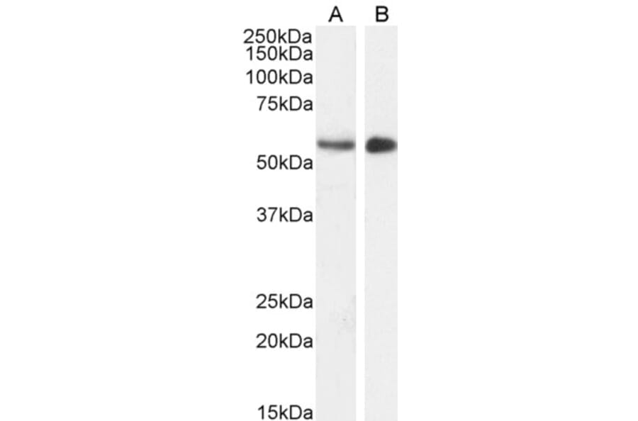
STK35 expression in Mouse (A) and Pig (B) Testes lysates analyzed by western blot. Cells were lysed in RIPA buffer and 35µg protein was run per lane. Primary incubation was performed with Anti-STK35 Antibody (A84004) at 1µg/ml (A) or 2µg/ml (B) and detected by chemiluminescence.
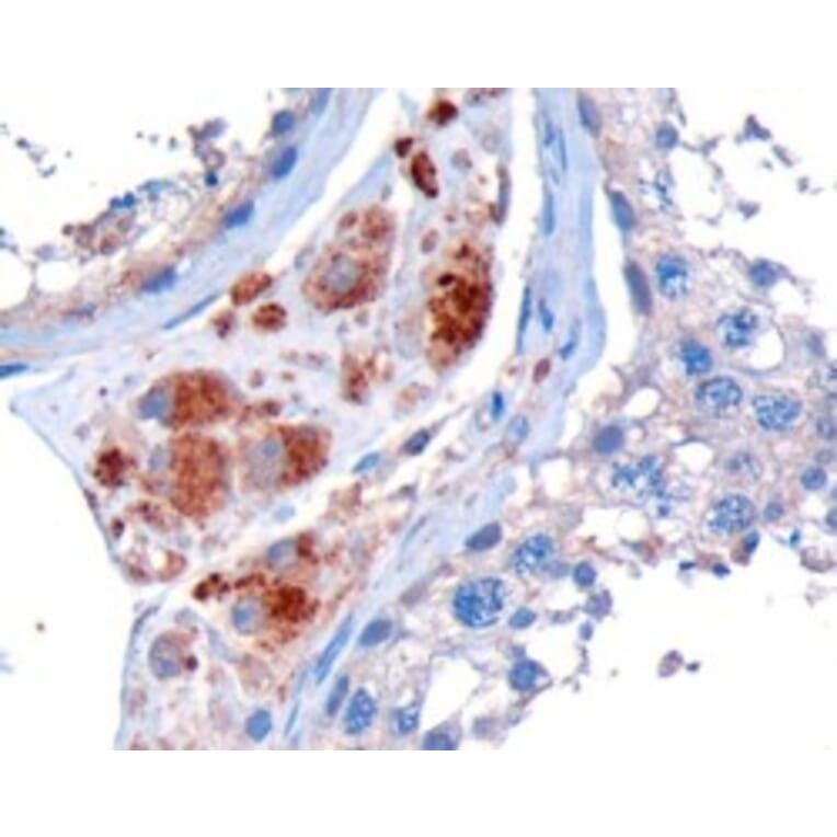
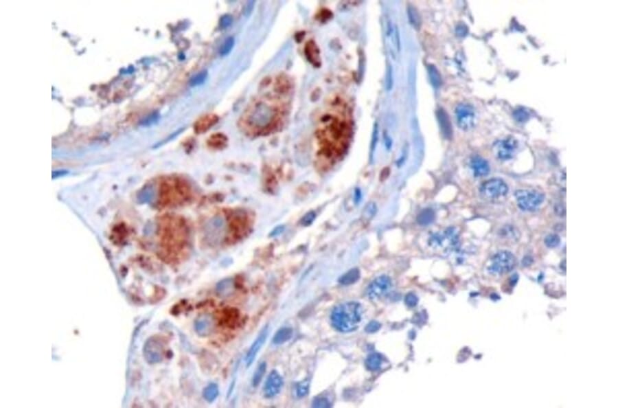
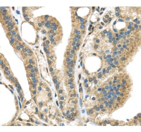
STK35 expression in Human Testis analyzed by immunohistochemistry. Tissue was paraffin-embedded, and antigen retrieval was achieved by microwaving in Tris/EDTA buffer, pH 9. Staining was performed with Anti-STK35 Antibody (A84004) at 10µg/ml and revealed with horseradish peroxidase (HRP).
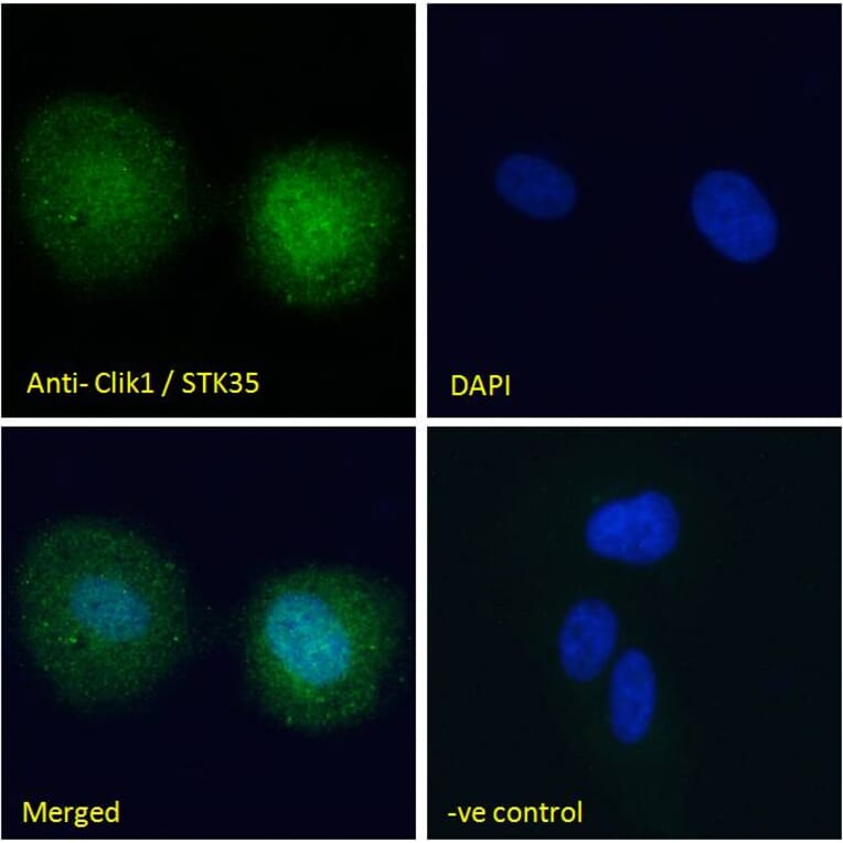
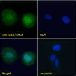
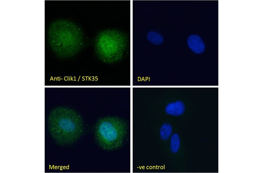
STK35 expression in U2OS cells analyzed by immunofluorescence. Cells were permeabilized with 0.15% Triton. Staining was performed with Anti-STK35 Antibody (A84004) at 10µg/ml for 1 hour and Alexa Fluor 488 secondary antibody at 2µg/ml. Nuclear staining shown and nuclei were stained with DAPI (blue). Negative control: Goat IgG Isotype Control at 10µg/ml followed by Alexa Fluor 488 secondary antibody at 2µg/ml.
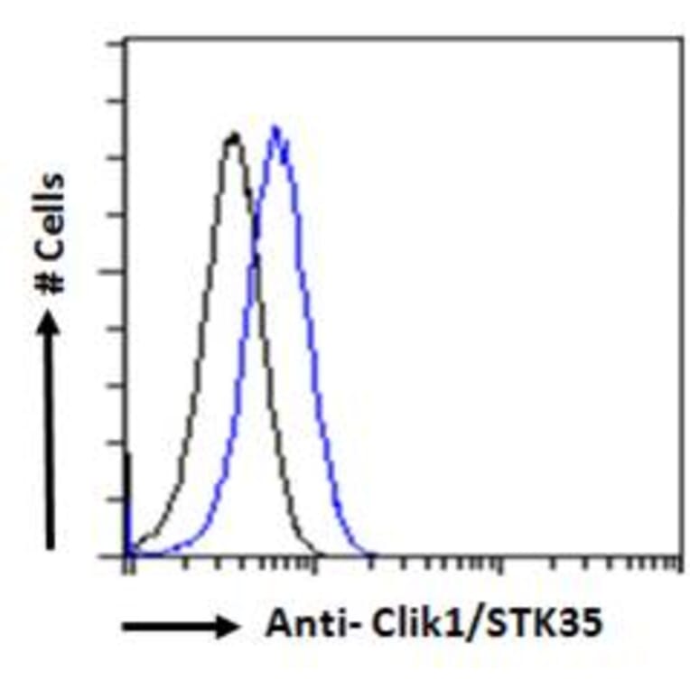
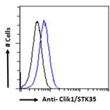
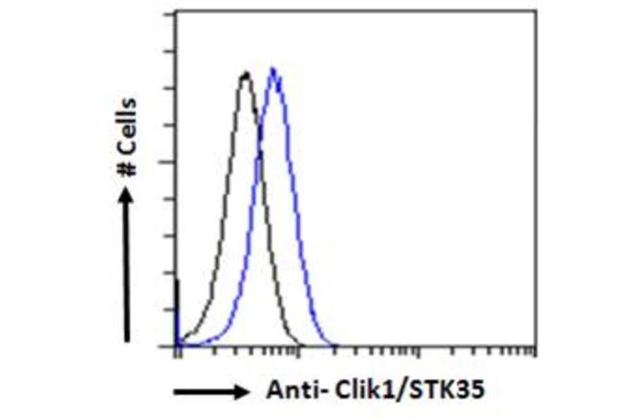
STK35 expression in HEK293 cells (blue line) analyzed by flow cytometry. Cells were fixed in PFA and permeabilized with 0.5% Triton. Staining was performed with Anti-STK35 Antibody (A84004) at 10µg/ml for 1 hour and Alexa Fluor 488 secondary antibody at 1µg/ml. Negative Control: Goat IgG Isotype Control (black line) followed by Alexa Fluor 488 secondary antibody.