TIMs are type I transmembrane glycoproteins with one Ig-like V-type domain and a Ser/Thr-rich mucin stalk. TIM-3 is expressed on the surface of effector T cells (CD4+Th1 and CD8+Tc1) but not on helper T cells (CD4+Th2 and CD8+Tc2). In chronic inflammation, autoimmune disorders, and some cancers, TIM-3 is upregulated on several other hematopoietic cell types. The Ig domain of TIM-3 interacts with a ligand on resting but not activated Th1 and Th2 cells. The glycosylated Ig domain of TIM-3 binds cell-associated galectin-9. This induces TIM-3 Tyr phosphorylation and pro-apoptotic signaling. TIM-3 functions as a negative regulator of Th1 cell activity. Its blockade results in increased IFN-gamma production, Th1 cell proliferation and cytotoxicity, regulatory T cell development, and increases in macrophage and neutrophil infiltration into sites of inflammation.
Applications
ELISA, IHC-P
Dilutions
IHC-P: 1-2 µg/ml
Reactivity
Human
Immunogen
Recombinant fragment, around amino acids 22-202, of human TIM3 protein. The exact sequence is proprietary.
Host
Mouse
Clonality
Monoclonal
Clone ID
TIM3/3113
Isotype
IgG2a
Light Chains
kappa
Conjugate
Unconjugated
Purification
Protein A/G chromatography.
Concentration
200 µg/ml
Molecular Weight
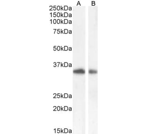
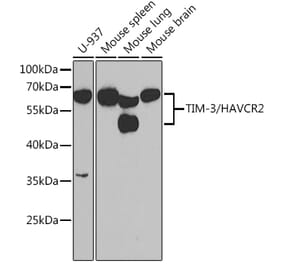
60 kDa
Product Form
Liquid
Formulation
Supplied in 10mM Phosphate Buffered Saline with 0.05% BSA and 0.05% Sodium Azide.
Storage
Shipped at 4°C. Upon delivery aliquot and store at -20°C. Avoid freeze / thaw cycles.
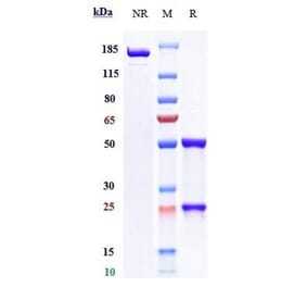
SDS-PAGE analysis of Anti-TIM3 Antibody [TIM3/3113] under non-reduced and reduced conditions; showing intact IgG and intact heavy and light chains, respectively. SDS-PAGE analysis confirms the integrity and purity of the antibody.
Analysis of protein array containing more than 19,000 full-length human proteins using Anti-TIM3 Antibody [TIM3/3113]. Z-Score and S- Score: The Z-score represents the strength of a signal that a monoclonal antibody (MAb) (in combination with a fluorescently-tagged anti-IgG secondary antibody) produces when binding to a particular protein on the HuProtTM array. Z-scores are described in units of standard deviations (SD's) above the mean value of all signals generated on that array. If targets on HuProtTM are arranged in descending order of the Z-score, the S-score is the difference (also in units of SD's) between the Z-score. S-score therefore represents the relative target specificity of a MAb to its intended target; a MAb is considered to be specific to its intended target, if the MAb has an S-score of at least 2.5. For example, if a MAb binds to protein X with a Z-score of 43 and to protein Y with a Z-score of 14, then the S-score for the binding of that MAb to protein X is equal to 29.


![Immunohistochemistry - Anti-TIM3 Antibody [TIM3/3113] (A251418) - Antibodies.com](https://cdn.antibodies.com/image/catalog/250/A250422_1.jpg?profile=product_top)
![Immunohistochemistry - Anti-TIM3 Antibody [TIM3/3113] (A250422) - Antibodies.com](https://cdn.antibodies.com/image/catalog/250/A250422_2.jpg?profile=product_top)
![Immunohistochemistry - Anti-TIM3 Antibody [TIM3/3113] (A253601) - Antibodies.com](https://cdn.antibodies.com/image/catalog/250/A250422_3.jpg?profile=product_top)
![SDS-PAGE - Anti-TIM3 Antibody [TIM3/3113] (A253601) - Antibodies.com](https://cdn.antibodies.com/image/catalog/250/A250422_4.jpg?profile=product_top)
![Protein Array - Anti-TIM3 Antibody [TIM3/3113] (A251611) - Antibodies.com](https://cdn.antibodies.com/image/catalog/250/A250422_5.jpg?profile=product_top)
![Immunohistochemistry - Anti-TIM3 Antibody [TIM3/3113] (A251418) - Antibodies.com](https://cdn.antibodies.com/image/catalog/250/A250422_1.jpg?profile=product_top_thumb)
![Immunohistochemistry - Anti-TIM3 Antibody [TIM3/3113] (A250422) - Antibodies.com](https://cdn.antibodies.com/image/catalog/250/A250422_2.jpg?profile=product_top_thumb)
![Immunohistochemistry - Anti-TIM3 Antibody [TIM3/3113] (A253601) - Antibodies.com](https://cdn.antibodies.com/image/catalog/250/A250422_3.jpg?profile=product_top_thumb)
![SDS-PAGE - Anti-TIM3 Antibody [TIM3/3113] (A253601) - Antibodies.com](https://cdn.antibodies.com/image/catalog/250/A250422_4.jpg?profile=product_top_thumb)
![Protein Array - Anti-TIM3 Antibody [TIM3/3113] (A251611) - Antibodies.com](https://cdn.antibodies.com/image/catalog/250/A250422_5.jpg?profile=product_top_thumb)
![Immunohistochemistry - Anti-TIM3 Antibody [TIM3/3113] (A251418) - Antibodies.com](https://cdn.antibodies.com/image/catalog/250/A250422_1.jpg?profile=product_image)
![Immunohistochemistry - Anti-TIM3 Antibody [TIM3/3113] (A250422) - Antibodies.com](https://cdn.antibodies.com/image/catalog/250/A250422_2.jpg?profile=product_image)
![Immunohistochemistry - Anti-TIM3 Antibody [TIM3/3113] (A253601) - Antibodies.com](https://cdn.antibodies.com/image/catalog/250/A250422_3.jpg?profile=product_image)
![SDS-PAGE - Anti-TIM3 Antibody [TIM3/3113] (A253601) - Antibodies.com](https://cdn.antibodies.com/image/catalog/250/A250422_4.jpg?profile=product_image)
![Protein Array - Anti-TIM3 Antibody [TIM3/3113] (A251611) - Antibodies.com](https://cdn.antibodies.com/image/catalog/250/A250422_5.jpg?profile=product_image)
![Immunohistochemistry - Anti-TIM3 Antibody [TIM3/4025] (A253577) - Antibodies.com](https://cdn.antibodies.com/image/catalog/250/A250423_1.jpg?profile=product_alternative)
![Immunohistochemistry - Anti-TIM3 Antibody [TIM3/4028] (A253603) - Antibodies.com](https://cdn.antibodies.com/image/catalog/250/A250424_1.jpg?profile=product_alternative)
![Immunohistochemistry - Anti-TIM3 Antibody [TIM3/4029] (A253604) - Antibodies.com](https://cdn.antibodies.com/image/catalog/250/A250425_1.jpg?profile=product_alternative)
![Immunohistochemistry - Anti-TIM3 Antibody [TIM3/4029] - BSA and Azide free (A250425) - Antibodies.com](https://cdn.antibodies.com/image/catalog/253/A253605_1.jpg?profile=product_alternative)
![Immunohistochemistry - Anti-TIM3 Antibody [TIM3/4028] - BSA and Azide free (A250424) - Antibodies.com](https://cdn.antibodies.com/image/catalog/253/A253604_1.jpg?profile=product_alternative)
![Immunohistochemistry - Anti-TIM3 Antibody [TIM3/4025] - BSA and Azide free (A250423) - Antibodies.com](https://cdn.antibodies.com/image/catalog/253/A253603_1.jpg?profile=product_alternative)
![Immunohistochemistry - Anti-TIM3 Antibody [TIM3/3113] - BSA and Azide free (A250422) - Antibodies.com](https://cdn.antibodies.com/image/catalog/253/A253602_1.jpg?profile=product_alternative)
![Western Blot - Anti-TIM3 Antibody [TIM3/4024] (A277833) - Antibodies.com](https://cdn.antibodies.com/image/catalog/277/A277833_1.jpg?profile=product_alternative)
![Immunohistochemistry - Anti-TIM3 Antibody [TIM3/4031] (A253599) - Antibodies.com](https://cdn.antibodies.com/image/catalog/250/A250420_1.jpg?profile=product_alternative)