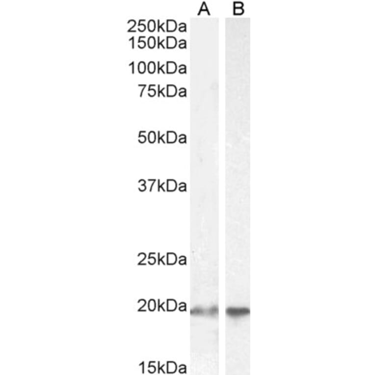
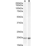
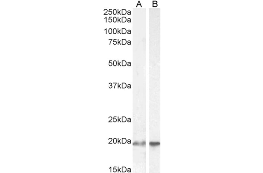
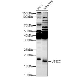
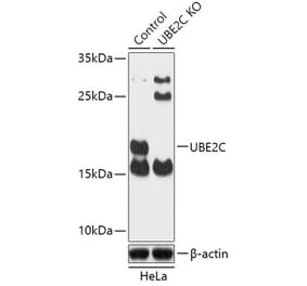
UBE2C expression in HEK293 (A) and HeLa (B) cell lysate analyzed by western blot. Cells were lysed in RIPA buffer and 35µg protein was run per lane. Primary antibody incubation was performed with Anti-UBE2C Antibody (A83441) at 2µg/ml and detected by chemiluminescence.
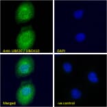
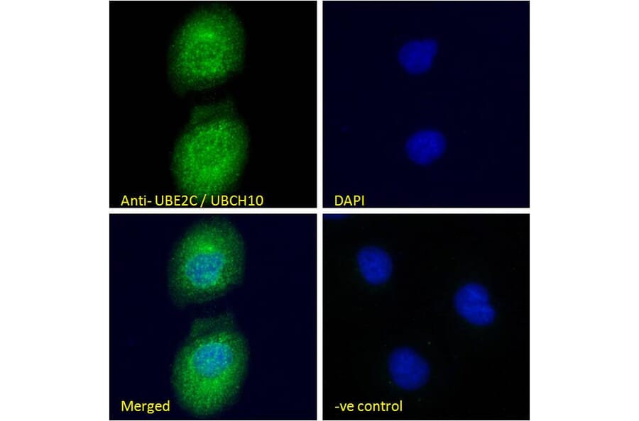
UBE2C expression in U2OS cells analyzed by immunofluorescence. Cells were permeabilized with 0.15% Triton. Staining was performed with Anti-UBE2C Antibody (A83441) at 10µg/ml for 1 hour and Alexa Fluor 488 secondary antibody at 4µg/ml. Cytoplasmic/plasma membrane staining shown and nuclei were stained with DAPI (blue). Negative control: Goat IgG Isotype Control at 10µg/ml followed by Alexa Fluor 488 secondary antibody at 4µg/ml.
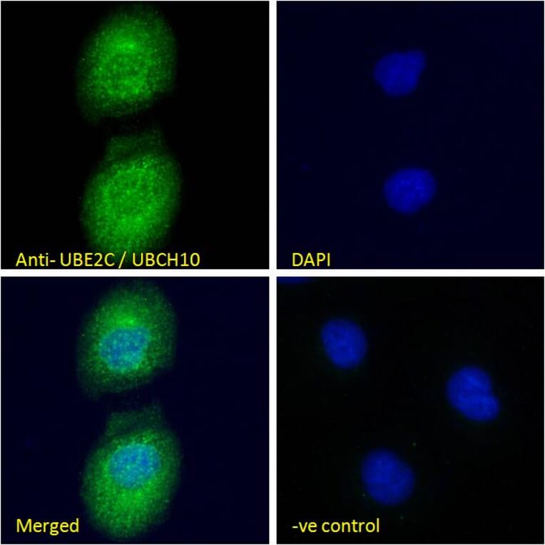
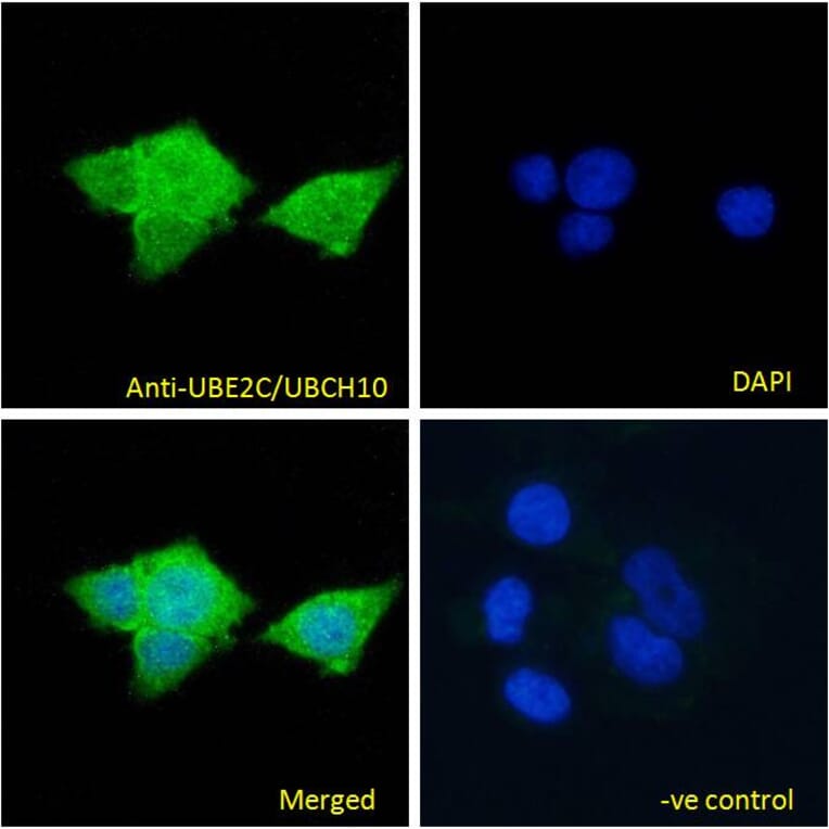
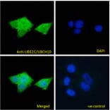
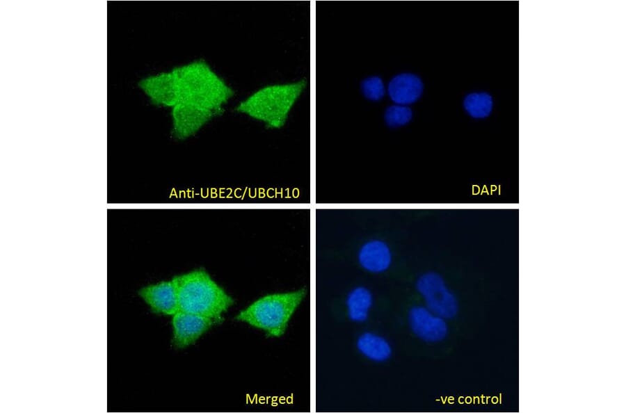
UBE2C expression in MCF7 cells analyzed by immunofluorescence. Cells were permeabilized with 0.15% Triton. Staining was performed with Anti-UBE2C Antibody (A83441) at 10µg/ml for 1 hour and Alexa Fluor 488 secondary antibody at 4µg/ml. Cytoplasmic staining shown and nuclei were stained with DAPI (blue). Negative control: Goat IgG Isotype Control at 10µg/ml followed by Alexa Fluor 488 secondary antibody at 4µg/ml.
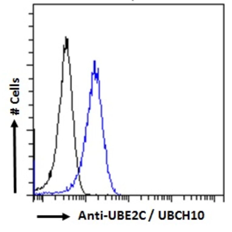
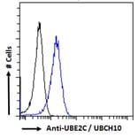
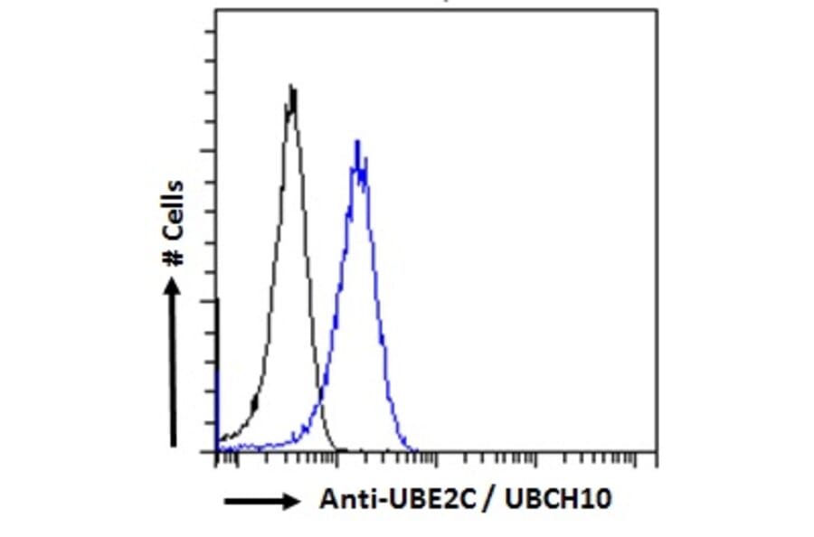
UBE2C expression in HeLa cells (blue line) analyzed by flow cytometry. Cells were fixed in PFA and permeabilized with 0.5% Triton. Staining was performed with Anti-UBE2C Antibody (A83441) at 10µg/ml for 1 hour and Alexa Fluor 488 secondary antibody at 2µg/ml. Negative Control: Goat IgG Isotype Control (black line) followed by Alexa Fluor 488 secondary antibody.
Publishing research using Anti-UBE2C Antibody (A83441)? Please let us know so that we can list the citation on this page.
Alternative products to Anti-UBE2C Antibody (A83441)


![Immunohistochemistry - Anti-UBE2C Antibody [CPTC-UBE2C-1] - BSA and Azide free (A248187) - Antibodies.com](https://cdn.antibodies.com/image/catalog/251/A251370_1.jpg?profile=product_alternative)
![Immunohistochemistry - Anti-UBE2C Antibody [CPTC-UBE2C-1] (A252050) - Antibodies.com](https://cdn.antibodies.com/image/catalog/248/A248187_1.jpg?profile=product_alternative)