Synthetic peptide corresponding to the internal region of human xCT.
Sequence
C-KGQTQNFKDAFSGRD
Host
Goat
Clonality
Polyclonal
Isotype
IgG
Conjugate
Unconjugated
Purification
Purified from goat serum by ammonium sulphate precipitation followed by antigen affinity chromatography using the immunizing peptide.
Concentration
500 µg/ml
Product Form
Liquid
Formulation
Supplied in Tris Buffered Saline, pH 7.3, with 0.5% BSA and 0.02% Sodium Azide.
Storage
Shipped at 4°C. Upon delivery aliquot and store at -20°C. Avoid freeze/thaw cycles.
Synonyms
Amino acid transport system xc-, Calcium channel blocker resistance protein CCBR1, Cystine/glutamate transporter, SLC7A11, Solute carrier family 7 member 11
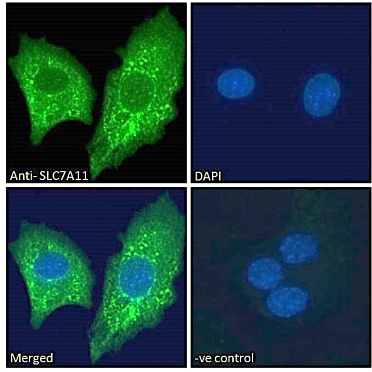
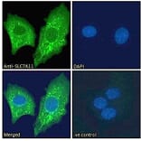
xCT expression in HepG2 cells analyzed by immunofluorescence. Cells were permeabilized with 0.15% Triton. Staining was performed with Anti-xCT Antibody (A84265) at 10µg/ml for 1 hour and Alexa Fluor 488 secondary antibody at 2µg/ml. Vesicle staining shown and nuclei were stained with DAPI (blue). Negative control: Goat IgG Isotype Control at 10µg/ml followed by Alexa Fluor 488 secondary antibody at 2µg/ml.
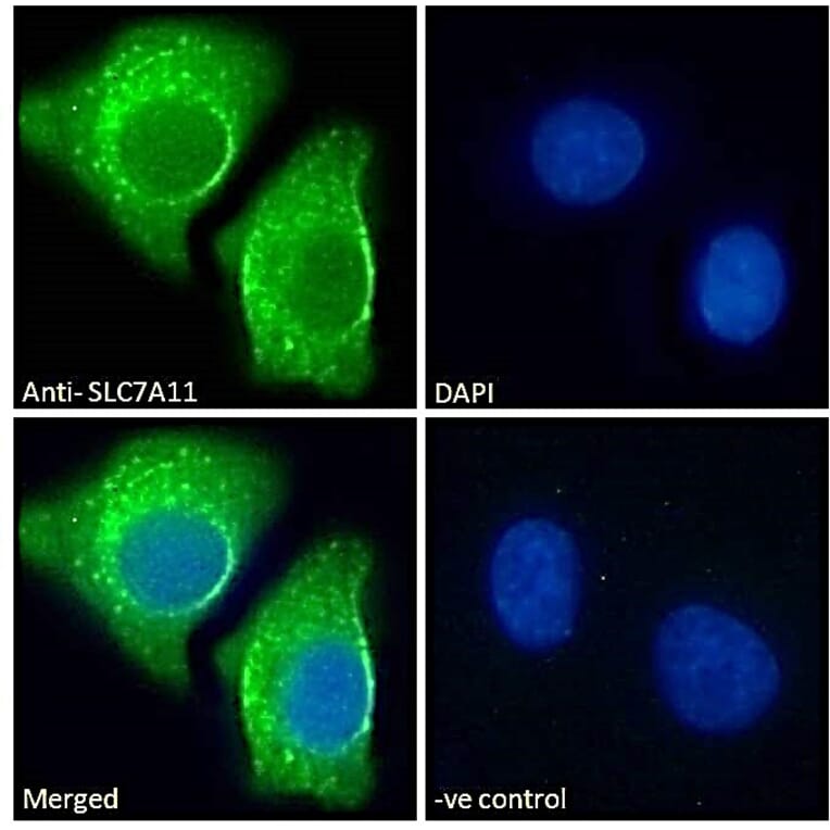
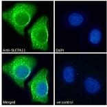
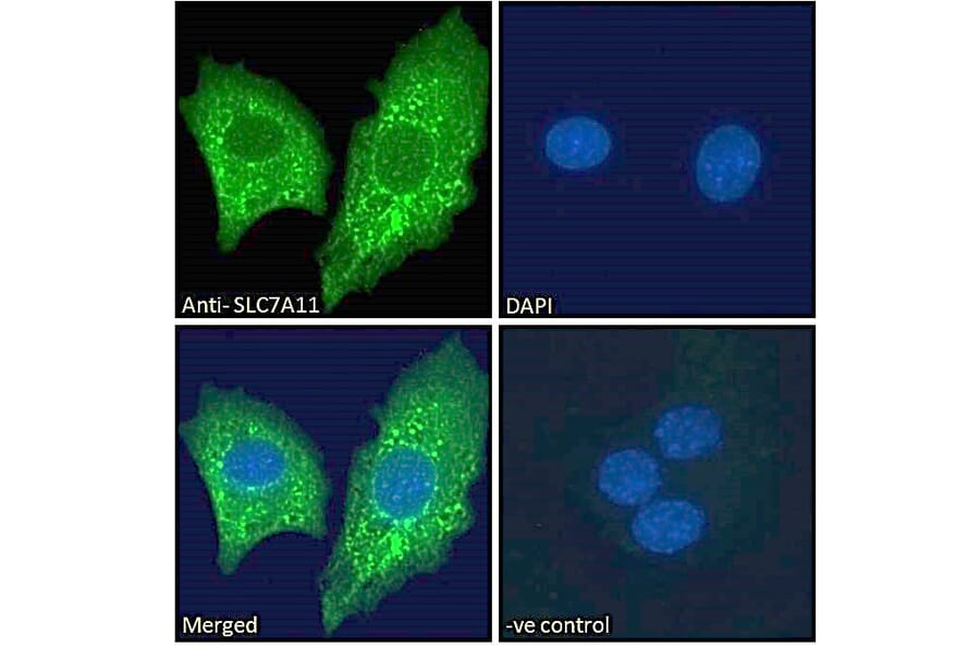
xCT expression in A549 cells analyzed by immunofluorescence. Cells were permeabilized with 0.15% Triton. Staining was performed with Anti-xCT Antibody (A84265) at 10µg/ml for 1 hour and Alexa Fluor 488 secondary antibody at 2µg/ml. ER/vesicle staining shown and nuclei were stained with DAPI (blue). Negative control: Goat IgG Isotype Control at 10µg/ml followed by Alexa Fluor 488 secondary antibody at 2µg/ml.
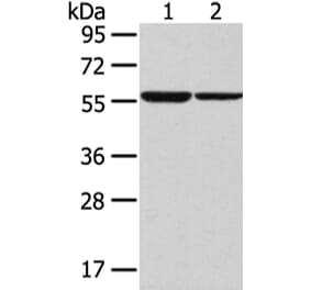
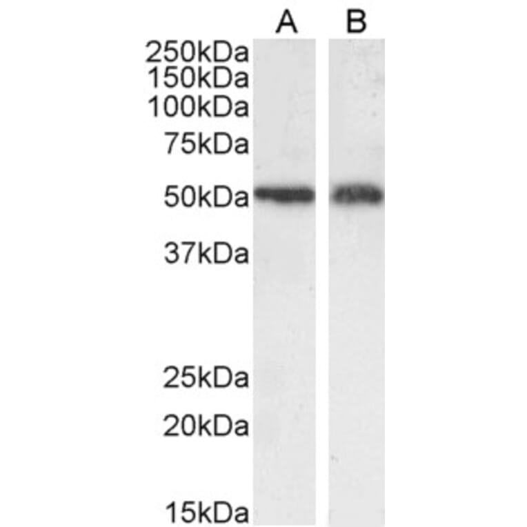
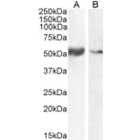
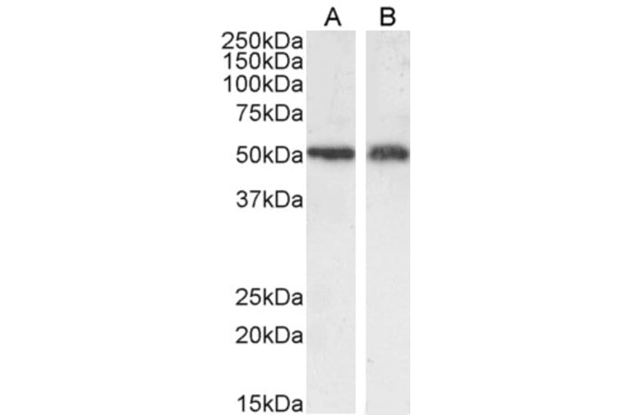
xCT expression in Human Smooth Muscle (A) and Tonsil (B) lysates analyzed by western blot. Cells were lysed in RIPA buffer and 35µg protein was run per lane. Primary antibody incubation was performed with Anti-xCT Antibody (A84265) at 0.1µg/ml and detected by chemiluminescence.
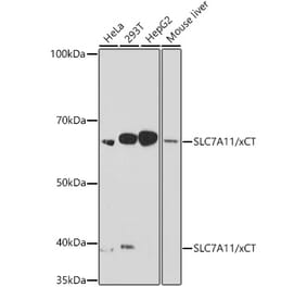
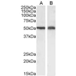
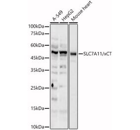
xCT expression in A549 (A) and U2OS (B) cell lysate analyzed by western blot. Cells were lysed in RIPA buffer and 35µg protein was run per lane. Primary antibody incubation was performed with Anti-xCT Antibody (A84265) at 0.3µg/ml and detected by chemiluminescence.
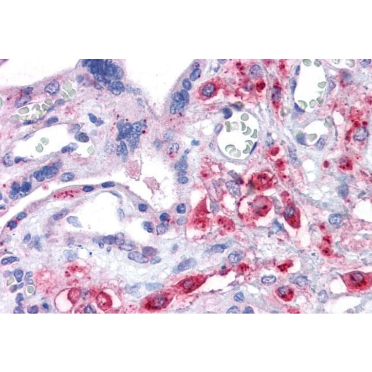
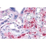
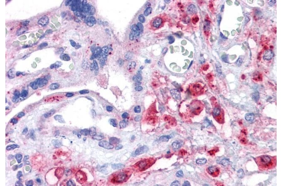
xCT expression in Human Placenta analyzed by immunohistochemistry. Tissue was paraffin-embedded, and antigen retrieval was achieved by steaming in citrate buffer, pH 6. Staining was performed with Anti-xCT Antibody (A84265) at 3.75µg/ml and revealed with alkaline phosphatase (AP). Note this data is from a previous batch and is not on sale.
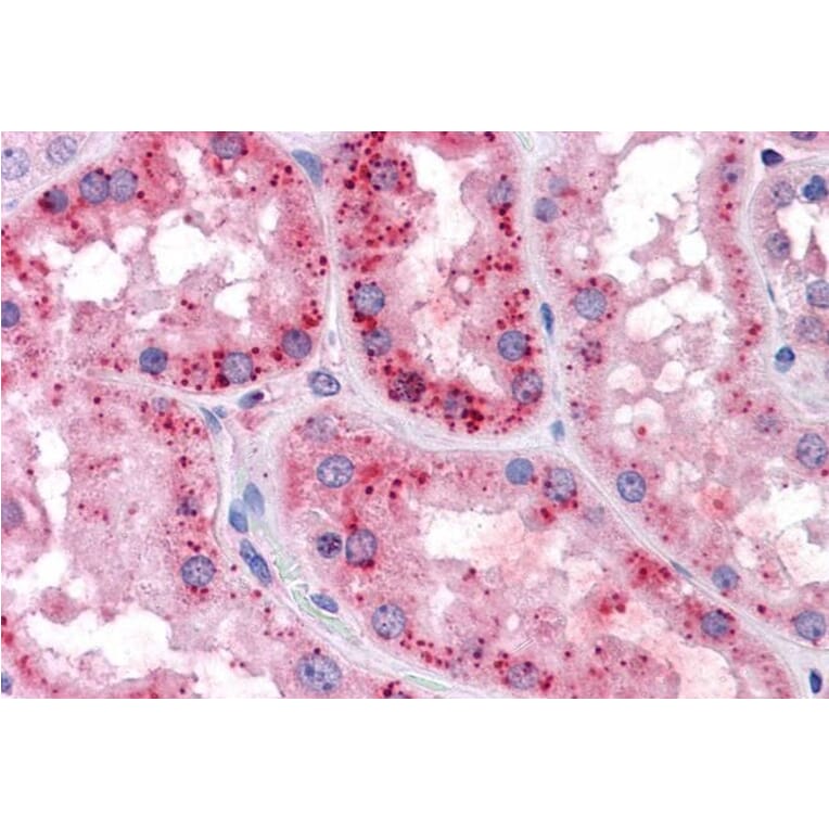
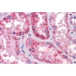
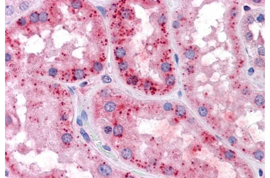
xCT expression in Human Kidney analyzed by immunohistochemistry. Tissue was paraffin-embedded, and antigen retrieval was achieved by steaming in citrate buffer, pH 6. Staining was performed with Anti-xCT Antibody (A84265) at 3.75µg/ml and revealed with alkaline phosphatase (AP). Note this data is from a previous batch and is not on sale.
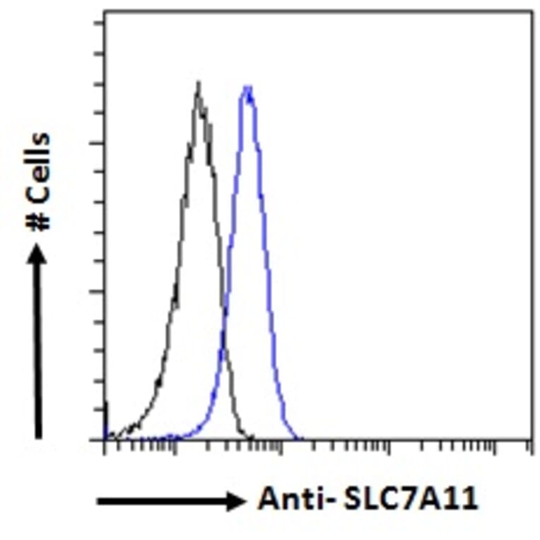
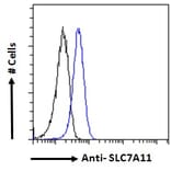
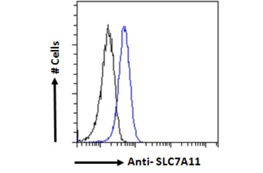
xCT expression in A549 cells (blue line) analyzed by flow cytometry. Cells were fixed in PFA and permeabilized with 0.5% Triton. Staining was performed with Anti-xCT Antibody (A84265) at 10µg/ml for 1 hour and Alexa Fluor 488 secondary antibody at 1µg/ml. Negative Control: Goat IgG Isotype Control (black line) followed by Alexa Fluor 488 secondary antibody.


![SDS-PAGE - Anti-xCT Antibody [Research Grade Biosimilar] - Low endotoxin, Azide free (A323867) - Antibodies.com](https://cdn.antibodies.com/image/catalog/323/A323867_1.jpg?profile=product_alternative)