This antibody reacts with ZAP-70, a 70 kDa protein tyrosine kinase expressed in T and NK cells. ZAP-70 is a molecule susceptible to degradation. It is recommended to use freshly prepared cell lysates (protease inhibitors are essential) to avoid non-specific staining of degradation products.
Applications
WB, ICC, Flow Cytometry (Intracellular)
Dilutions
Flow Cytometry: Intracellular staining: 2-5 µg/ml; Positive Control: HPB-ALL human peripheral blood T cell leukemia cell line, WB: 0.5-2 µg/ml.
Reactivity
Human
Immunogen
Bacterially expressed fusion protein representing C-terminal part (160 amino acids) of human ZAP-70 protein with histidine tag.
Host
Mouse
Clonality
Monoclonal
Clone ID
ZAP-03
Isotype
IgG1
Conjugate
Unconjugated
Purification
Protein A chromatography.
Concentration
1 mg/ml
Predicted MW
70 kDa
Product Form
Liquid
Formulation
Supplied in Phosphate Buffered Saline, pH 7.4, with 15 mM Sodium Azide.
Storage
Shipped at 4°C. Upon delivery aliquot and store at -20°C. Avoid freeze/thaw cycles.
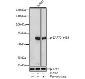
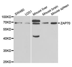
Western blotting analysis of human ZAP70 using Anti-ZAP70 Antibody [ZAP-03] (A85696) on lysates of Jurkat cell line and HEK-293T cell line (negative control) under reducing and non-reducing conditions. Nitrocellulose membrane was probed with 2 µg/ml of anti-ZAP70 mouse monoclonal antibody followed by IRDye800-conjugated anti-mouse secondary antibody. A specific band was detected for ZAP70 protein at approximately 70 kDa.


![WB - Anti-ZAP70 Antibody [ZAP-03] (A85696)](https://cdn.antibodies.com/image/catalog/85/A85696_1.jpg?profile=product_top)
![Flow Cytometry - Anti-ZAP70 Antibody [ZAP-03] (A85696)](https://cdn.antibodies.com/image/catalog/85/A85696_2.jpg?profile=product_top)
![Flow Cytometry - Anti-ZAP70 Antibody [ZAP-03] (A85696)](https://cdn.antibodies.com/image/catalog/85/A85696_3.jpg?profile=product_top)
![Flow Cytometry - Anti-ZAP70 Antibody [ZAP-03] (A85696)](https://cdn.antibodies.com/image/catalog/85/A85696_4.jpg?profile=product_top)
![WB - Anti-ZAP70 Antibody [ZAP-03] (A85696)](https://cdn.antibodies.com/image/catalog/85/A85696_1.jpg?profile=product_top_thumb)
![Flow Cytometry - Anti-ZAP70 Antibody [ZAP-03] (A85696)](https://cdn.antibodies.com/image/catalog/85/A85696_2.jpg?profile=product_top_thumb)
![Flow Cytometry - Anti-ZAP70 Antibody [ZAP-03] (A85696)](https://cdn.antibodies.com/image/catalog/85/A85696_3.jpg?profile=product_top_thumb)
![Flow Cytometry - Anti-ZAP70 Antibody [ZAP-03] (A85696)](https://cdn.antibodies.com/image/catalog/85/A85696_4.jpg?profile=product_top_thumb)
![WB - Anti-ZAP70 Antibody [ZAP-03] (A85696)](https://cdn.antibodies.com/image/catalog/85/A85696_1.jpg?profile=product_image)
![Flow Cytometry - Anti-ZAP70 Antibody [ZAP-03] (A85696)](https://cdn.antibodies.com/image/catalog/85/A85696_2.jpg?profile=product_image)
![Flow Cytometry - Anti-ZAP70 Antibody [ZAP-03] (A85696)](https://cdn.antibodies.com/image/catalog/85/A85696_3.jpg?profile=product_image)
![Flow Cytometry - Anti-ZAP70 Antibody [ZAP-03] (A85696)](https://cdn.antibodies.com/image/catalog/85/A85696_4.jpg?profile=product_image)
![Immunohistochemistry - Anti-ZAP70 Antibody [ZAP70/2047] - BSA and Azide free (A250346) - Antibodies.com](https://cdn.antibodies.com/image/catalog/253/A253526_1.jpg?profile=product_alternative)
![Immunohistochemistry - Anti-ZAP70 Antibody [ZAP70/2035] - BSA and Azide free (A250344) - Antibodies.com](https://cdn.antibodies.com/image/catalog/253/A253524_1.jpg?profile=product_alternative)
![Immunohistochemistry - Anti-ZAP70 Antibody [ZAP70/2047] (A253524) - Antibodies.com](https://cdn.antibodies.com/image/catalog/250/A250346_1.jpg?profile=product_alternative)
![Immunohistochemistry - Anti-ZAP70 Antibody [ZAP70/2035] (A253521) - Antibodies.com](https://cdn.antibodies.com/image/catalog/250/A250344_1.jpg?profile=product_alternative)
![Immunohistochemistry - Anti-ZAP70 Antibody [2F3.2] (A253501) - Antibodies.com](https://cdn.antibodies.com/image/catalog/250/A250340_1.jpg?profile=product_alternative)
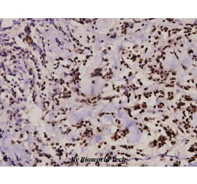
![Immunohistochemistry - Anti-ZAP70 Antibody [2F3.2] - BSA and Azide free (A250340) - Antibodies.com](https://cdn.antibodies.com/image/catalog/253/A253520_1.jpg?profile=product_alternative)