This product recognizes the heavy and light chains of Mouse IgG.
Applications
ICC/IF, WB, IHC-P, IHC-Fr
Recommended Dilutions
IF: 1-10 µg/mL, WB: 50-100 ng/mL, IHC: 1-10 µg/mL, . The optimal concentration should be determined by the end-user.
Clonality
Polyclonal
Isotype
IgG
Conjugate
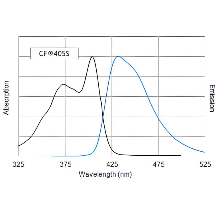
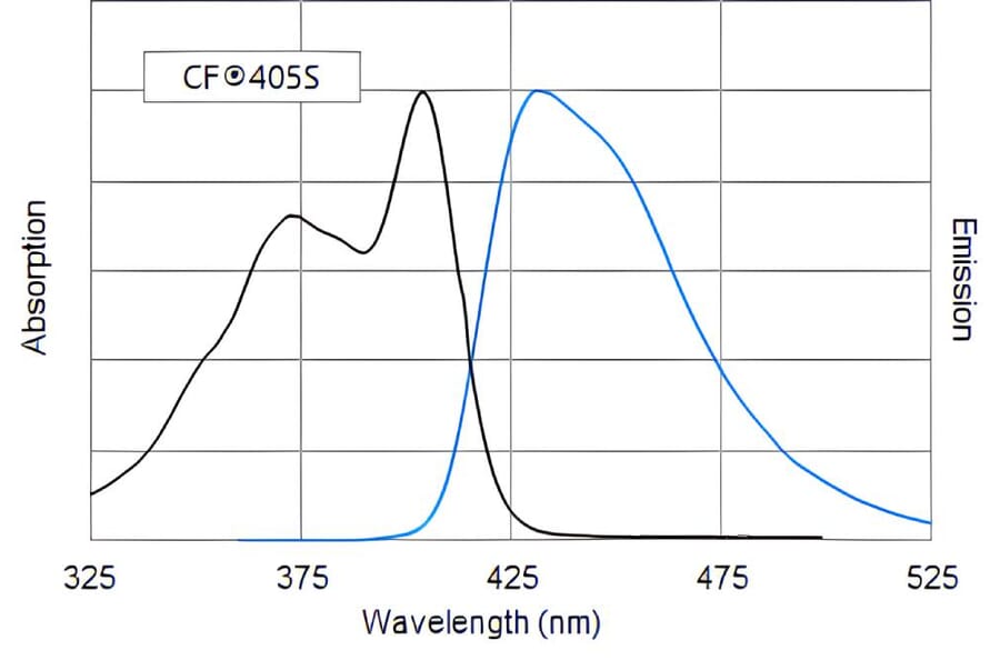
CF®405S
Product Form
Liquid
Concentration
2 mg/mL
Formulation
After reconstitution; phosphate Buffered Saline with 15 mg/ml BSA and 20 mg/ml trehalose.
Storage
Shipped at 4°C. Upon delivery aliquot and store at -20°C. Avoid freeze/thaw cycles. This product is also photosensitive and should be protected from light. Should this product contain a precipitate we recommend microcentrifugation before use. CF® Dyes are guaranteed for at least 6 months from data of receipt when stored correctly.
General Notes
Looking for a specific protein conjugate to simplify your workflow? We offer a library of over 2,000 targets conjugated to your choice of CF® dye. To enquire about a custom product, contact us directly.
Disclaimer
This product is for research use only. It is not intended for diagnostic or therapeutic use.
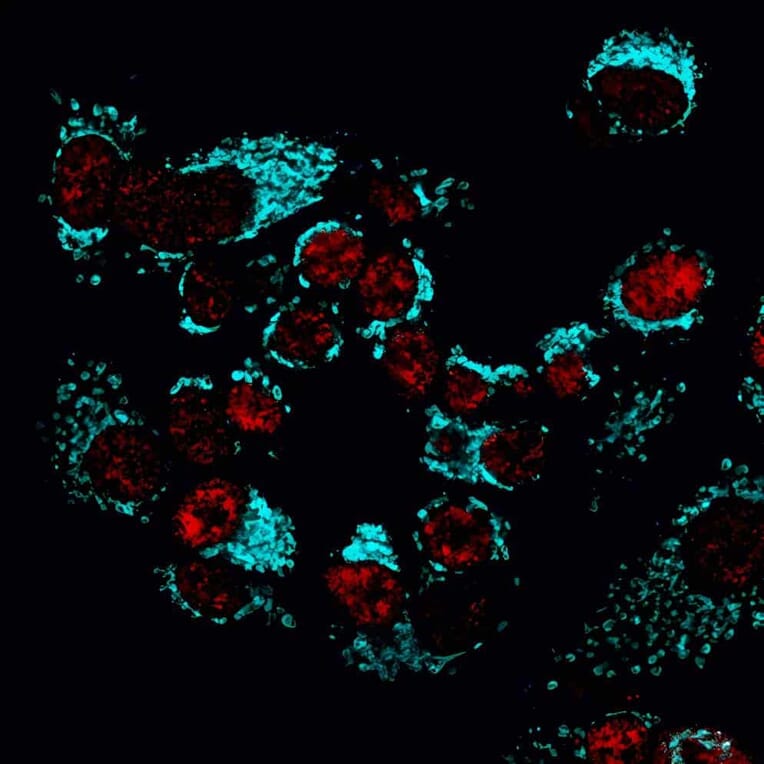
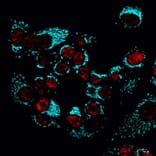
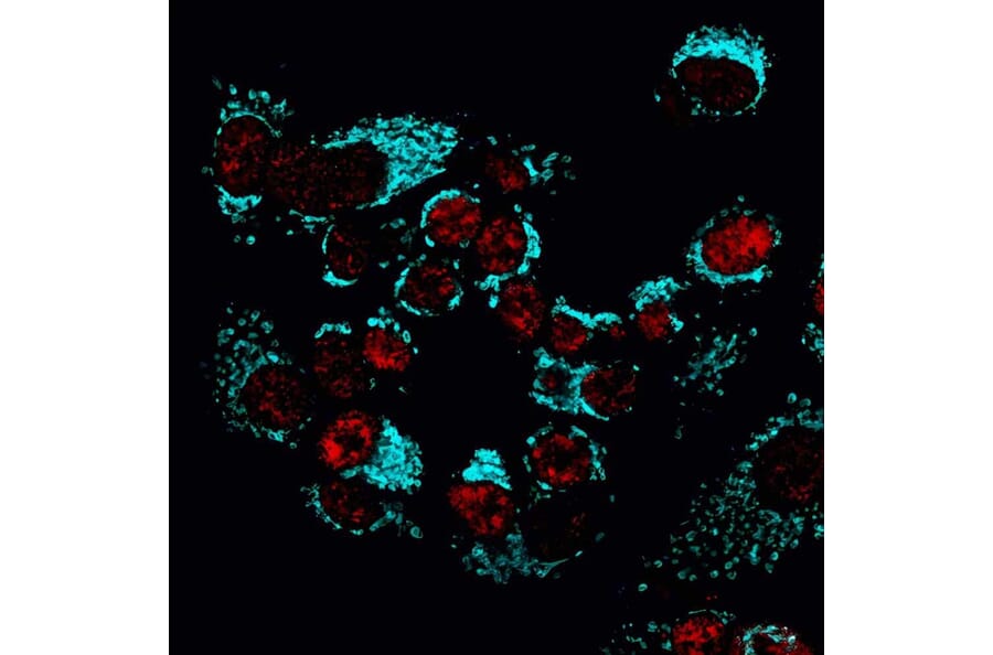
MCF-7 cells stained with rabbit anti-COX IV followed by CF®405S goat anti-rabbit to label mitochondria (blue), and mouse anti-phosphorylated RNA polymerase II followed by CF®647 goat anti-mouse to label nuclei (red).
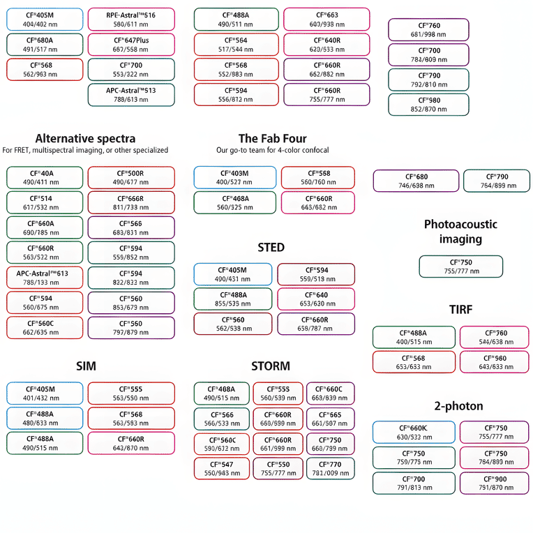
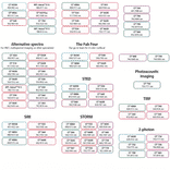
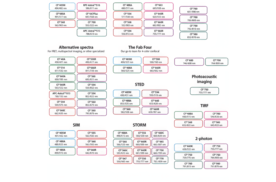
This chart summarizes commonly used CF® dyes grouped by their suitability for specific imaging modalities, including alternative spectra applications, four-color confocal imaging, near-infrared western blotting, photoacoustic imaging, STED, SIM, STORM, TIRF, and two-photon microscopy. Each dye is shown with its characteristic excitation and emission wavelengths (nm), providing a practical reference for selecting spectrally compatible dyes and optimizing multicolor experimental design across a range of fluorescence techniques.
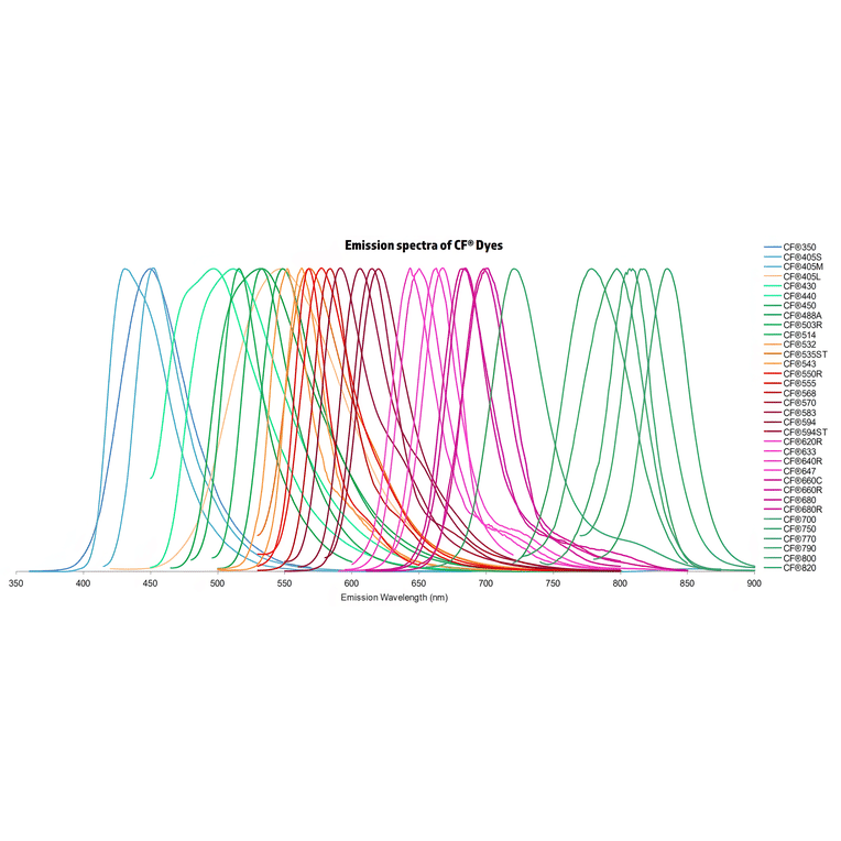
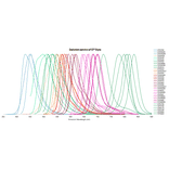
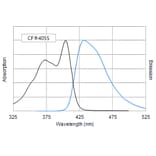
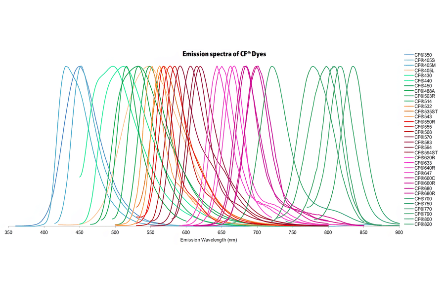
Normalized emission spectra of the CF® dye family spanning the visible to near-infrared range are shown, illustrating the spectral diversity and overlap between dyes. Curves represent relative fluorescence intensity as a function of emission wavelength (nm), with peak positions corresponding to each dye’s characteristic emission maximum. This reference highlights the broad coverage of CF® dyes for multicolor fluorescence applications and aids in selecting compatible dye combinations for imaging, flow cytometry, and other fluorescence-based assays.
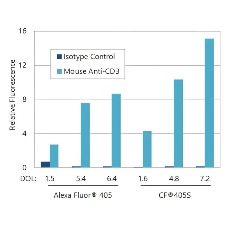
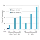
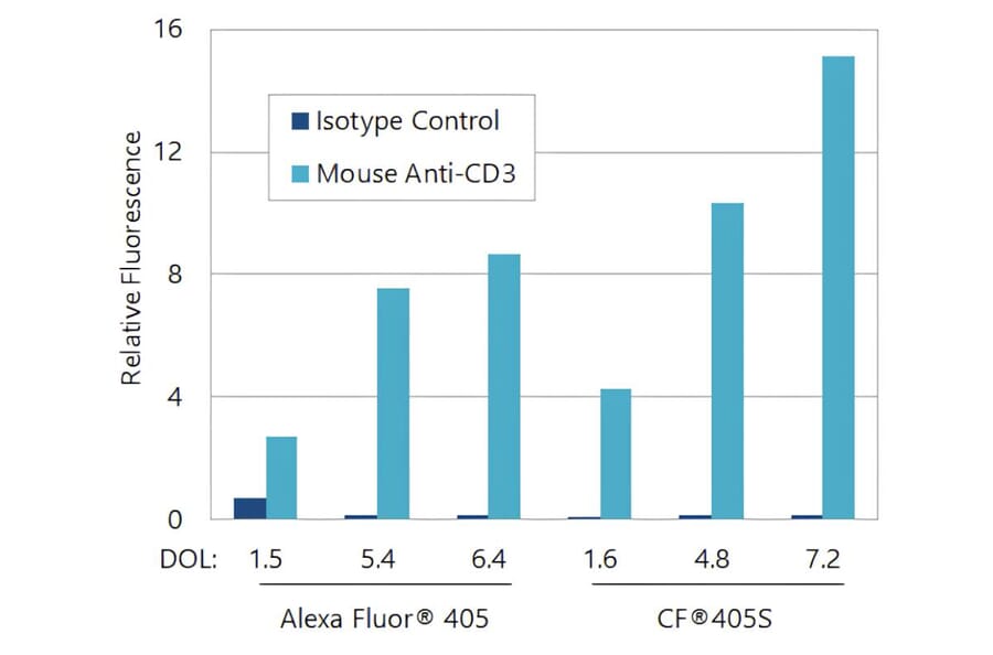
CF®405S produces brighter antibody conjugates than Alexa Fluor® 405. Flow cytometry analysis was performed in the Pacific Blue® detection channel on a BD LSRII cytometer using Jurkat cells stained with mouse anti-CD3 or an isotype control, followed by goat anti-mouse secondary antibodies conjugated to either Alexa Fluor® 405 or CF®405S at varying degrees of labeling. Bars represent geometric mean fluorescence intensity.
Publishing research using Goat Anti-Mouse IgG H&L Antibody (CF®405S) - Highly Cross-Adsorbed (A343988)? Please let us know so that we can list the citation on this page.