This product recognizes the heavy and light chains of Mouse IgG.
Applications
ICC/IF, WB, IHC-P, IHC-Fr
Recommended Dilutions
IF: 1-10 µg/mL, WB: 50-100 ng/mL, IHC: 1-10 µg/mL, . The optimal concentration should be determined by the end-user.
Clonality
Polyclonal
Isotype
IgG
Conjugate
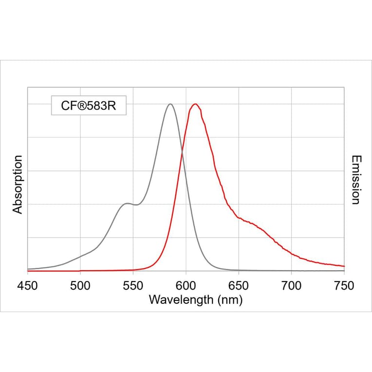
CF®583R
Product Form
Liquid
Concentration
2 mg/mL
Formulation
Supplied in Phosphate Buffered Saline with 2 mg/ml BSA and 0.05% Sodium Azide.
Storage
Shipped at 4°C. Upon delivery aliquot and store at -20°C. Avoid freeze/thaw cycles. This product is also photosensitive and should be protected from light. Should this product contain a precipitate we recommend microcentrifugation before use. CF® Dyes are guaranteed for at least 6 months from data of receipt when stored correctly.
General Notes
Looking for a specific protein conjugate to simplify your workflow? We offer a library of over 2,000 targets conjugated to your choice of CF® dye. To enquire about a custom product, contact us directly.
Disclaimer
This product is for research use only. It is not intended for diagnostic or therapeutic use.
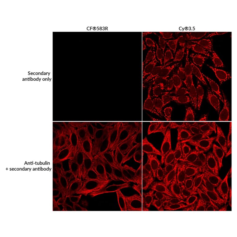
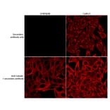
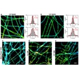
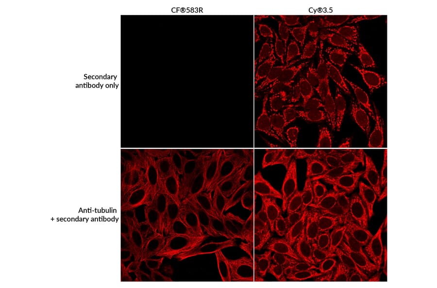
Because of its enhanced aqueous solubility, CF®583R produces markedly lower non-specific background staining than Cy®3.5. Methanol-fixed HeLa cells were incubated either with a mouse anti-tubulin primary antibody or with no primary antibody control, followed by goat anti-mouse secondary antibodies conjugated to CF®583R or Cy®3.5. In the absence of primary antibody, the Cy®3.5 conjugate generated pronounced background fluorescence, whereas background signal remained minimal with the CF®583R conjugate, highlighting its superior specificity.
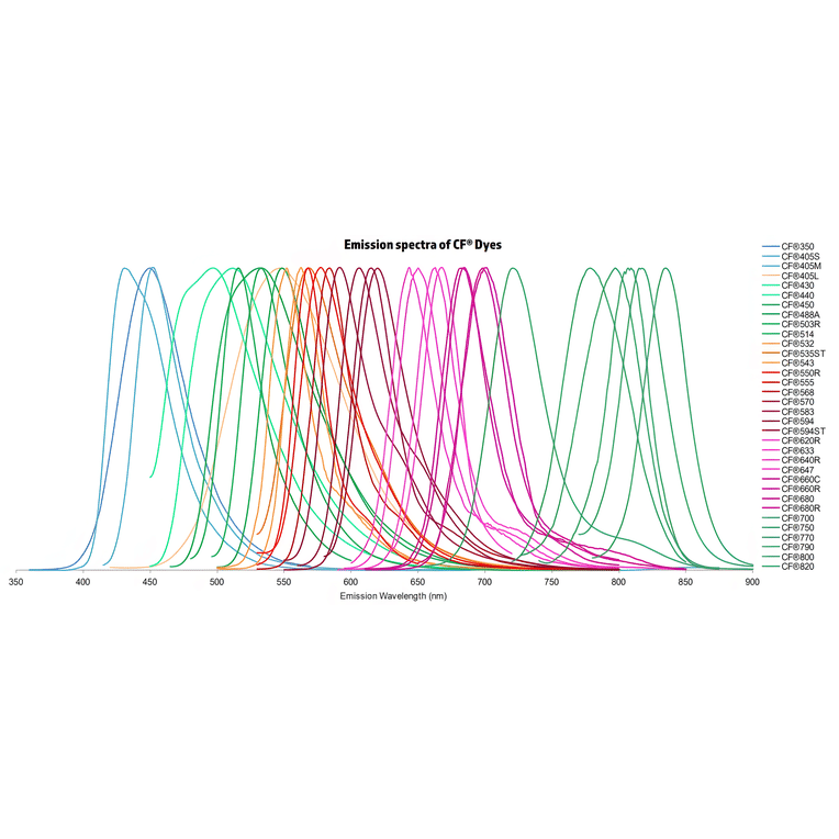
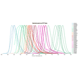
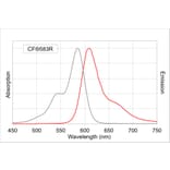
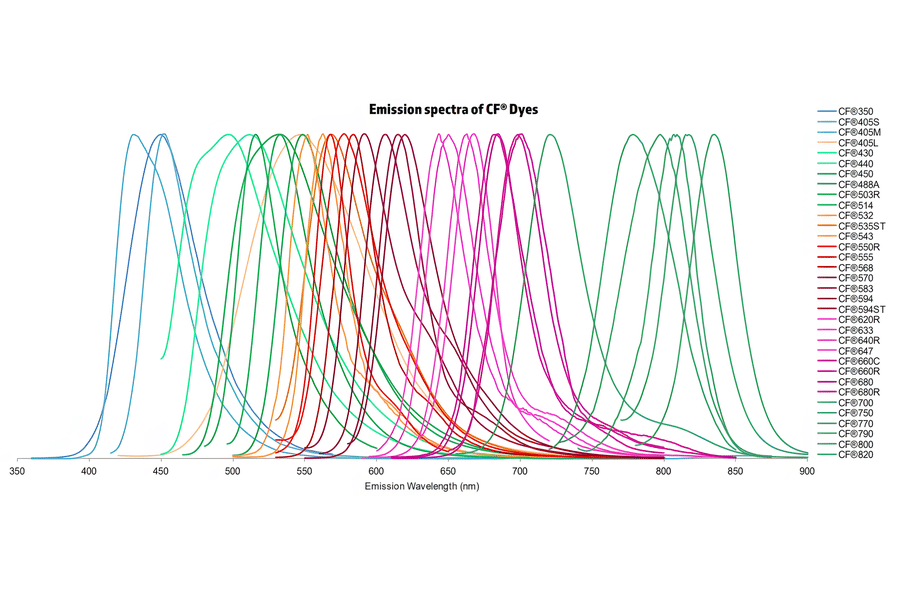
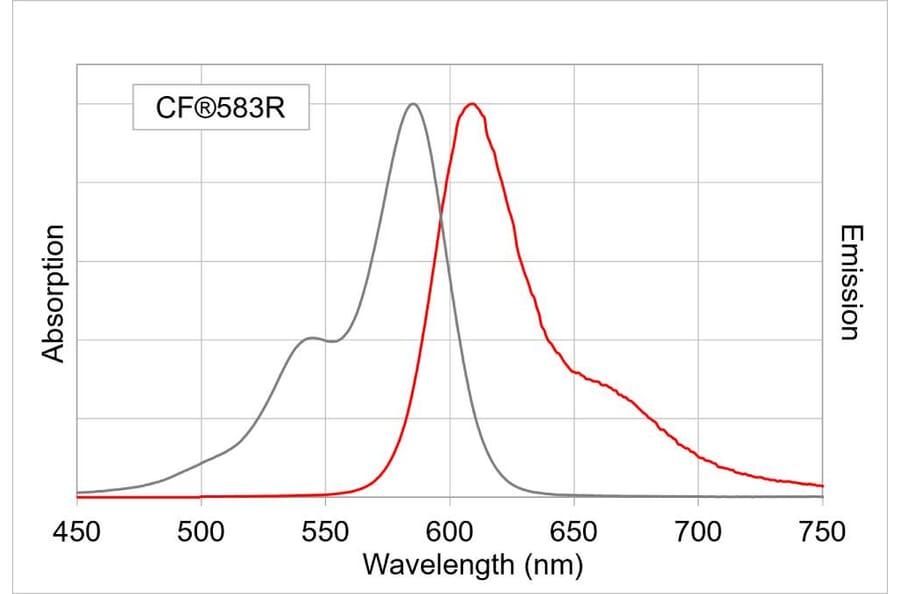
Normalized emission spectra of the CF® dye family spanning the visible to near-infrared range are shown, illustrating the spectral diversity and overlap between dyes. Curves represent relative fluorescence intensity as a function of emission wavelength (nm), with peak positions corresponding to each dye’s characteristic emission maximum. This reference highlights the broad coverage of CF® dyes for multicolor fluorescence applications and aids in selecting compatible dye combinations for imaging, flow cytometry, and other fluorescence-based assays.
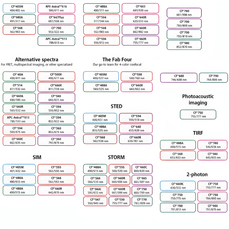
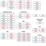
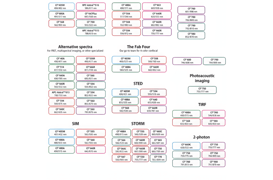
This chart summarizes commonly used CF® dyes grouped by their suitability for specific imaging modalities, including alternative spectra applications, four-color confocal imaging, near-infrared western blotting, photoacoustic imaging, STED, SIM, STORM, TIRF, and two-photon microscopy. Each dye is shown with its characteristic excitation and emission wavelengths (nm), providing a practical reference for selecting spectrally compatible dyes and optimizing multicolor experimental design across a range of fluorescence techniques.
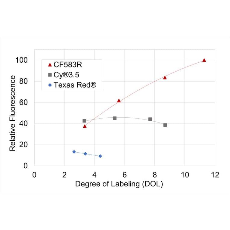
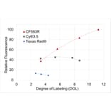
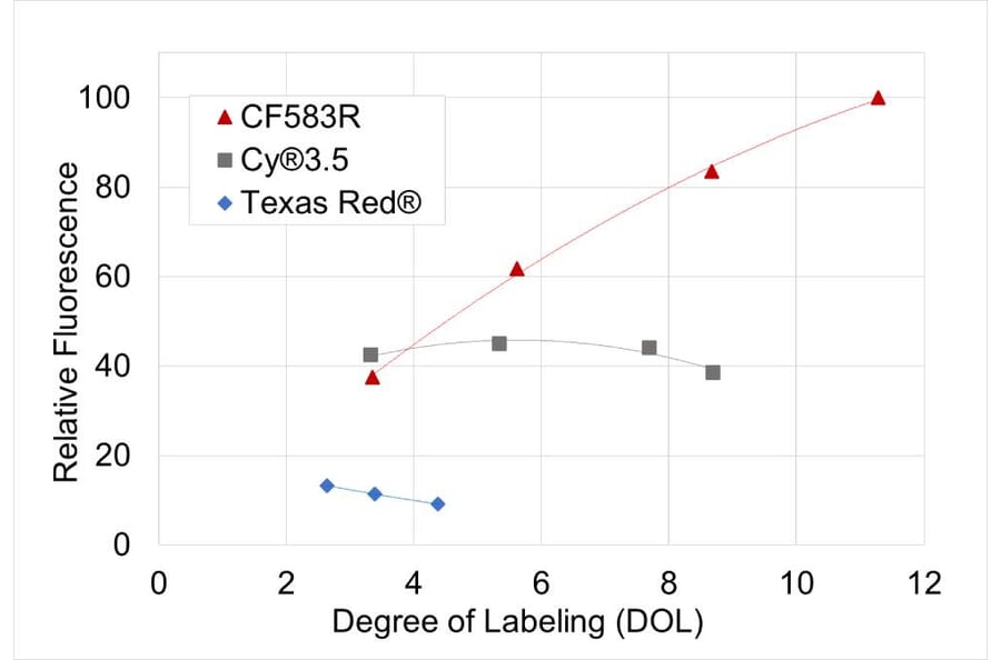
CF®583R yields brighter antibody conjugates at lower degrees of labeling than Cy®3.5 and Texas Red®. Shown is the relative fluorescence of goat anti-mouse conjugates bearing the indicated dyes across a range of labeling densities, expressed as the number of dye molecules per antibody.
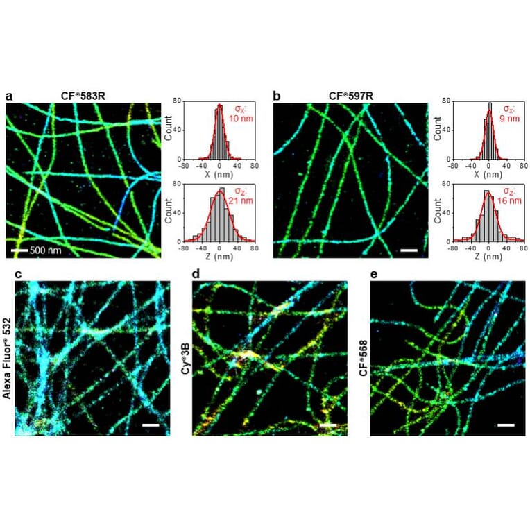
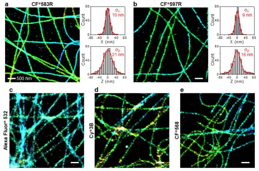
Comparison of representative (d)STORM images of microtubules in fixed COS-7 cells immunolabeled with different fluorescent dyes. (d)STORM imaging was performed under standard conditions using a tris-based imaging buffer containing 100 mM cysteamine and an oxygen-scavenging system. Microtubules were labeled with (a) CF®583R, (b) CF®597R, (c) Alexa Fluor® 532, (d) Cy3B, and (e) CF®568. Localization distributions for individual CF®583R and CF®597R molecules are shown in the X (in-plane, top) and Z (axial depth, bottom) directions. Gaussian fits (red curves) yield localization precisions of approximately 10 nm laterally and 20 nm axially. Color coding represents depth (Z) information. Images and analysis were provided courtesy of Bowen Wang, Michael Xiong, and Professor Ke Xu, College of Chemistry, University of California, Berkeley.
Publishing research using Goat Anti-Mouse IgG H&L Antibody (CF®583R) - Highly Cross-Adsorbed (A344060)? Please let us know so that we can list the citation on this page.