IF: 1-10 µg/mL, WB: 50-100 ng/mL, IHC: 1-10 µg/mL, . The optimal concentration should be determined by the end-user.
Clonality
Polyclonal
Isotype
IgG
Conjugate
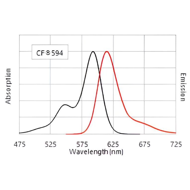
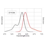
CF®594
Product Form
Liquid
Concentration
1 mg/mL
Formulation
Supplied in Phosphate Buffered Saline with 2 mg/ml BSA and 0.05% Sodium Azide.
Storage
Shipped at 4°C. Upon delivery aliquot and store at -20°C. Avoid freeze/thaw cycles. This product is also photosensitive and should be protected from light. Should this product contain a precipitate we recommend microcentrifugation before use. CF® Dyes are guaranteed for at least 6 months from data of receipt when stored correctly.
General Notes
Looking for a specific protein conjugate to simplify your workflow? We offer a library of over 2,000 targets conjugated to your choice of CF® dye. To enquire about a custom product, contact us directly.
Disclaimer
This product is for research use only. It is not intended for diagnostic or therapeutic use.
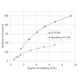
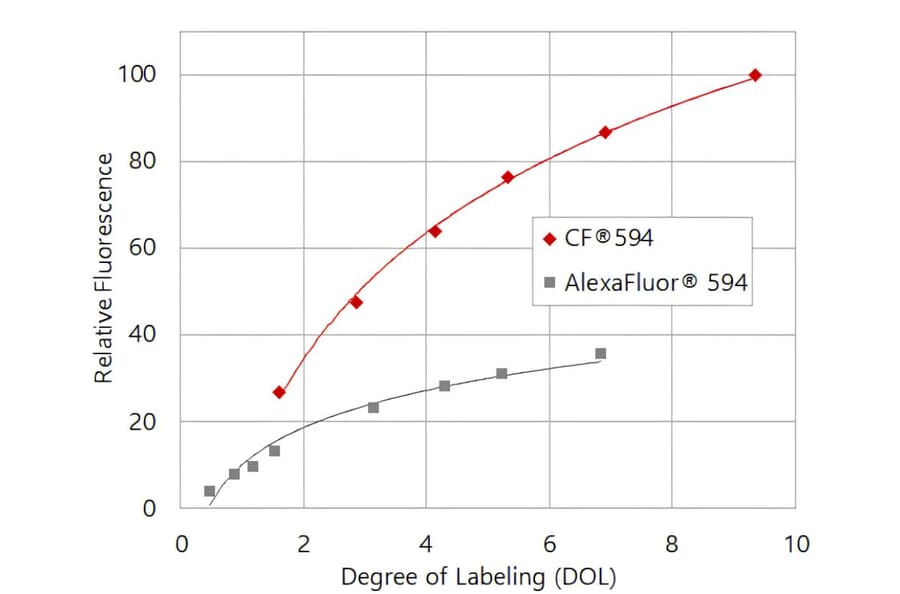
CF®594 produces brighter antibody conjugates than Alexa Fluor® 594. Relative fluorescence intensity of goat anti-mouse IgG conjugates is shown as a function of degree of labeling (DOL, dye molecules per antibody).
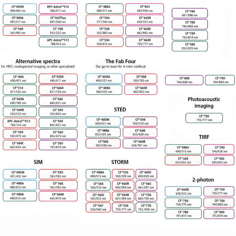
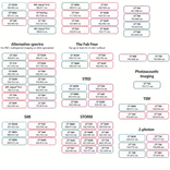
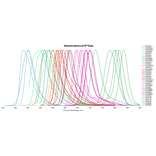
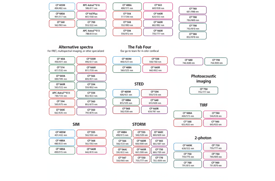
This chart summarizes commonly used CF® dyes grouped by their suitability for specific imaging modalities, including alternative spectra applications, four-color confocal imaging, near-infrared western blotting, photoacoustic imaging, STED, SIM, STORM, TIRF, and two-photon microscopy. Each dye is shown with its characteristic excitation and emission wavelengths (nm), providing a practical reference for selecting spectrally compatible dyes and optimizing multicolor experimental design across a range of fluorescence techniques.
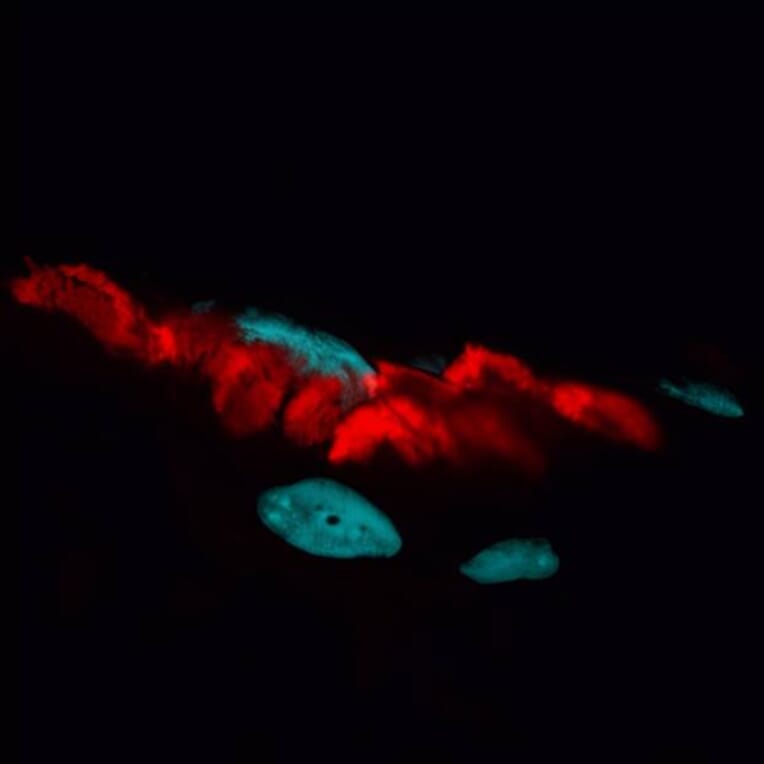
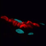
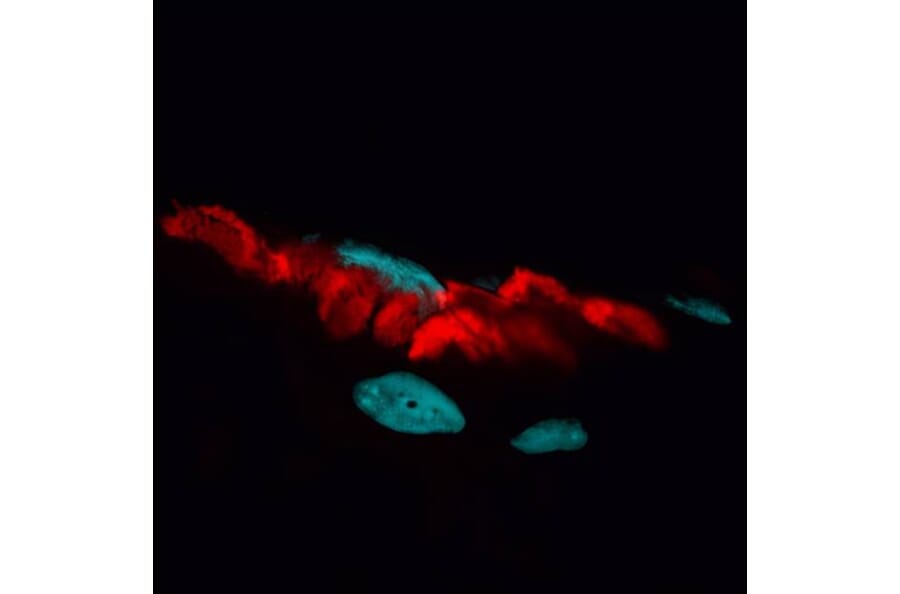
Neuromuscular junctions in a rat skeletal muscle section stained with CF®594 a-bungarotoxin to label acetylcholine receptors (red), with nuclei counterstained using DAPI (blue).
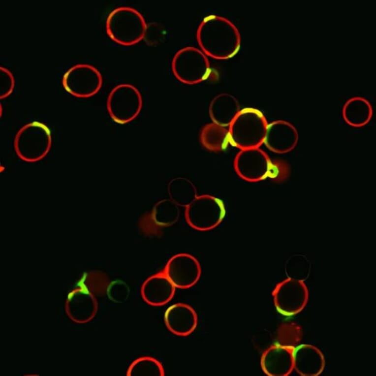
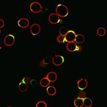
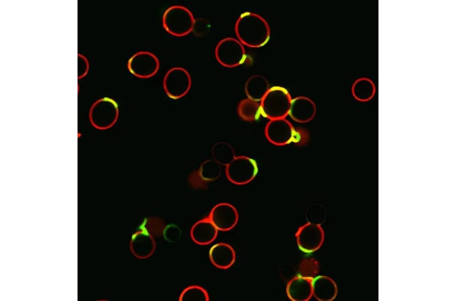
Saccharomyces cerevisiae yeast stained with CF®594 wheat germ agglutinin to label cell walls (red) and CF®488A concanavalin A to label bud scars (green).
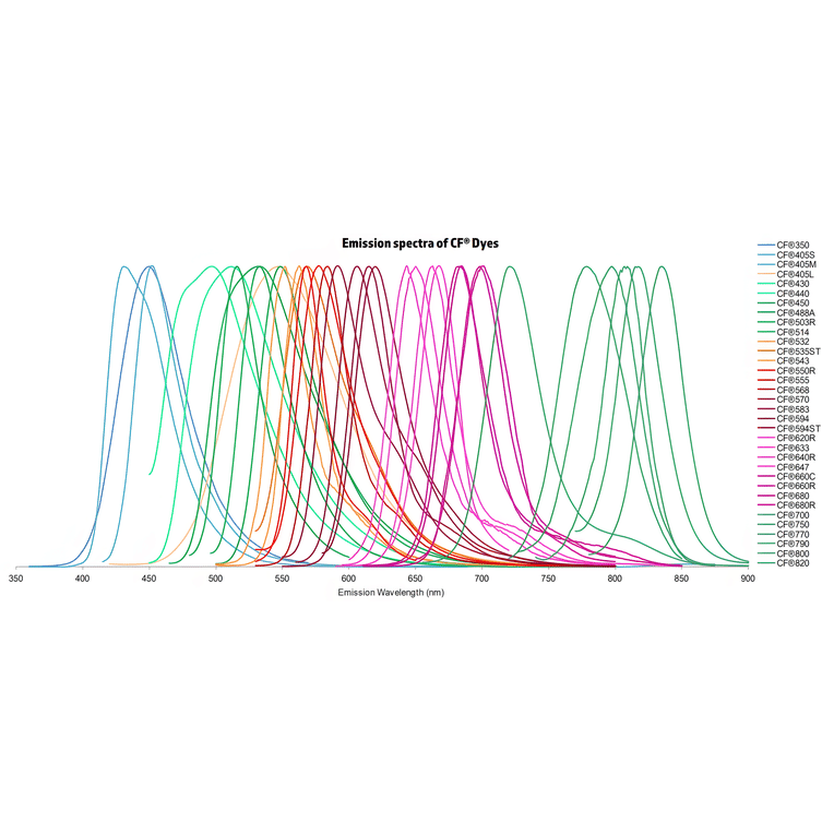
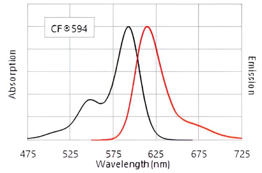
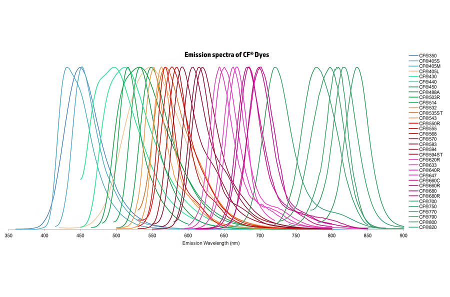
Normalized emission spectra of the CF® dye family spanning the visible to near-infrared range are shown, illustrating the spectral diversity and overlap between dyes. Curves represent relative fluorescence intensity as a function of emission wavelength (nm), with peak positions corresponding to each dye’s characteristic emission maximum. This reference highlights the broad coverage of CF® dyes for multicolor fluorescence applications and aids in selecting compatible dye combinations for imaging, flow cytometry, and other fluorescence-based assays.
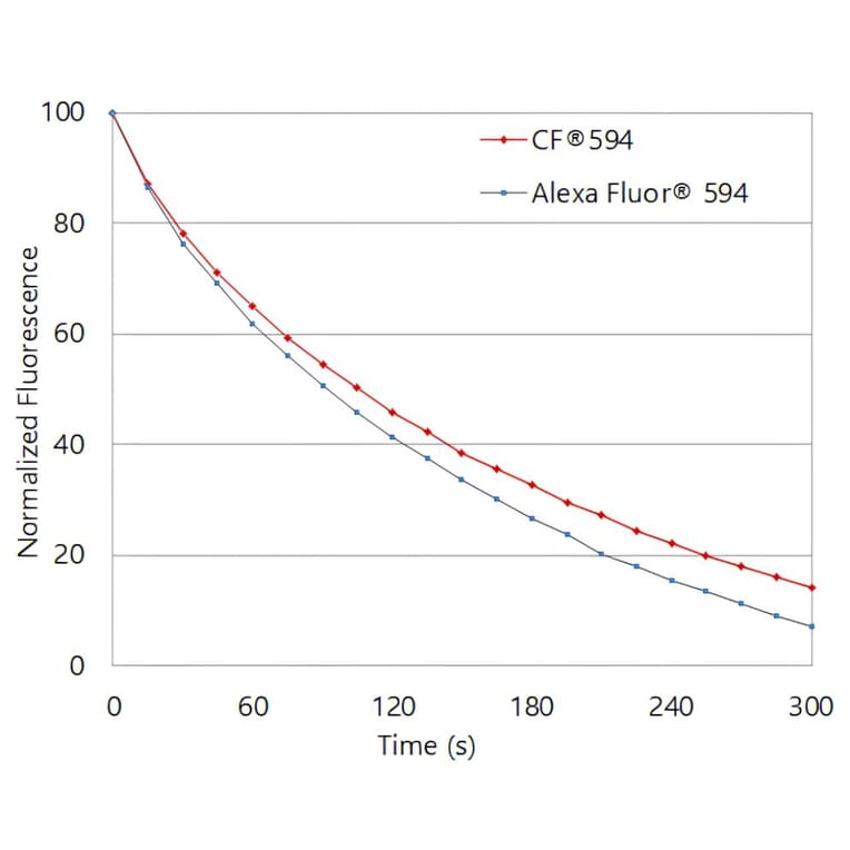
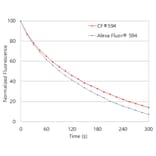
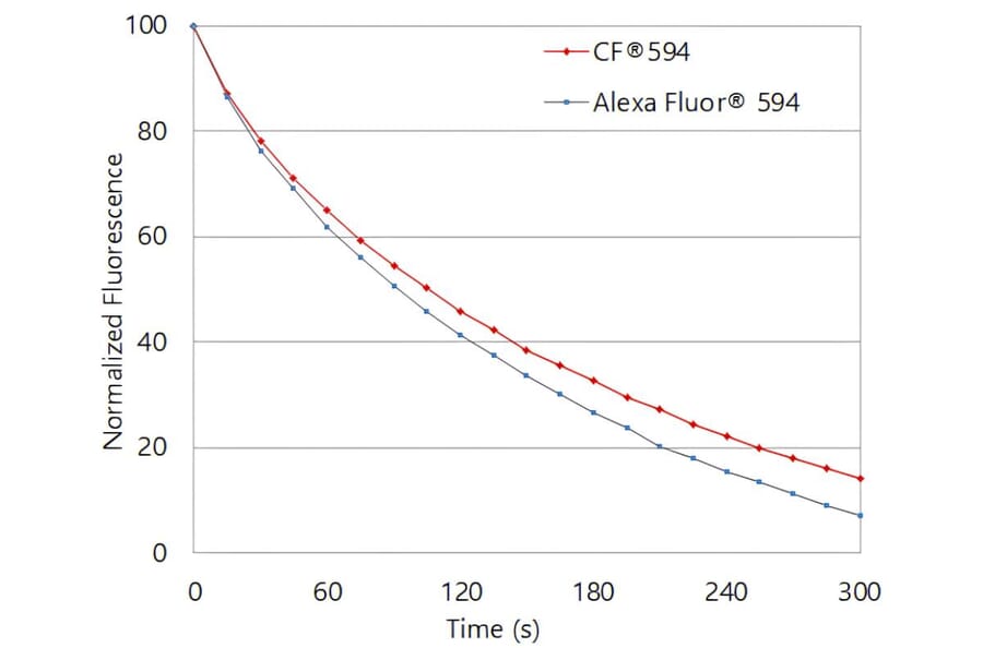
Photostability comparison of CF®594 and Alexa Fluor® 594. Jurkat cells were stained with mouse anti-CD3 primary antibody followed by CF®594 or Alexa Fluor® 594 goat anti-mouse IgG secondary antibodies. Cells were continuously illuminated, and fluorescence intensity was monitored over time.
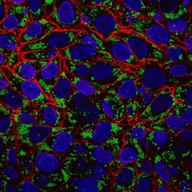
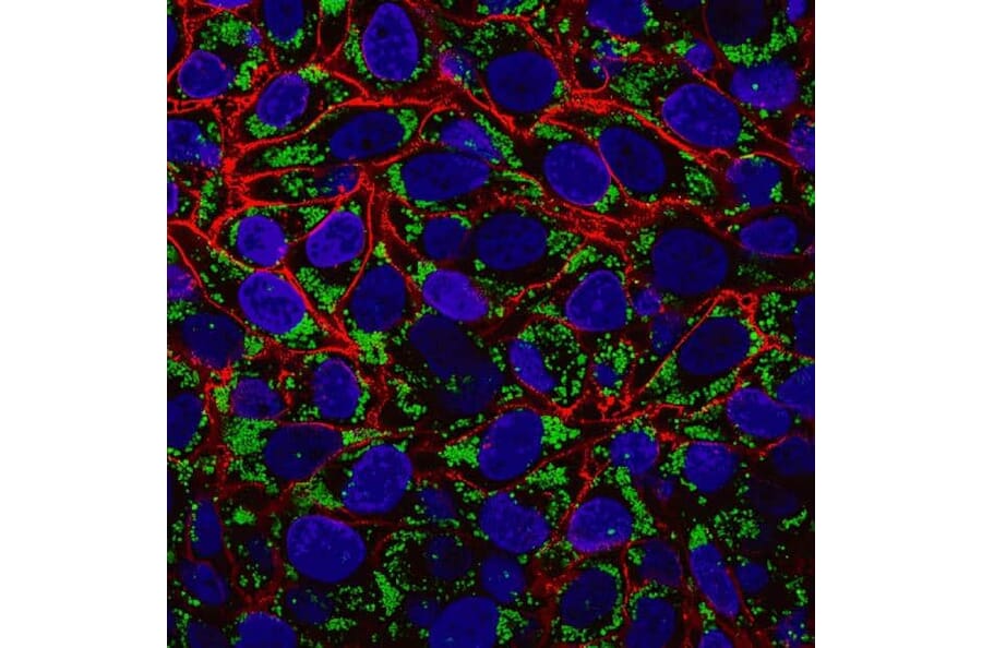
Fixed oleic acid–treated HeLa cells stained with LipidSpot™ 488 to visualize lipid droplets (green), CF®594 wheat germ agglutinin to label the cell surface (red), and Hoechst 33258 to stain nuclei (blue).
Publishing research using Goat F(ab')2 Anti-Alpaca IgG, VHH domain + Llama IgG, VHH domain Antibody (CF®594) - Highly Cross-Adsorbed (A344041)? Please let us know so that we can list the citation on this page.