IF: 1-10 µg/mL, WB: 50-100 ng/mL, IHC: 1-10 µg/mL, . The optimal concentration should be determined by the end-user.
Clonality
Polyclonal
Isotype
IgG
Conjugate
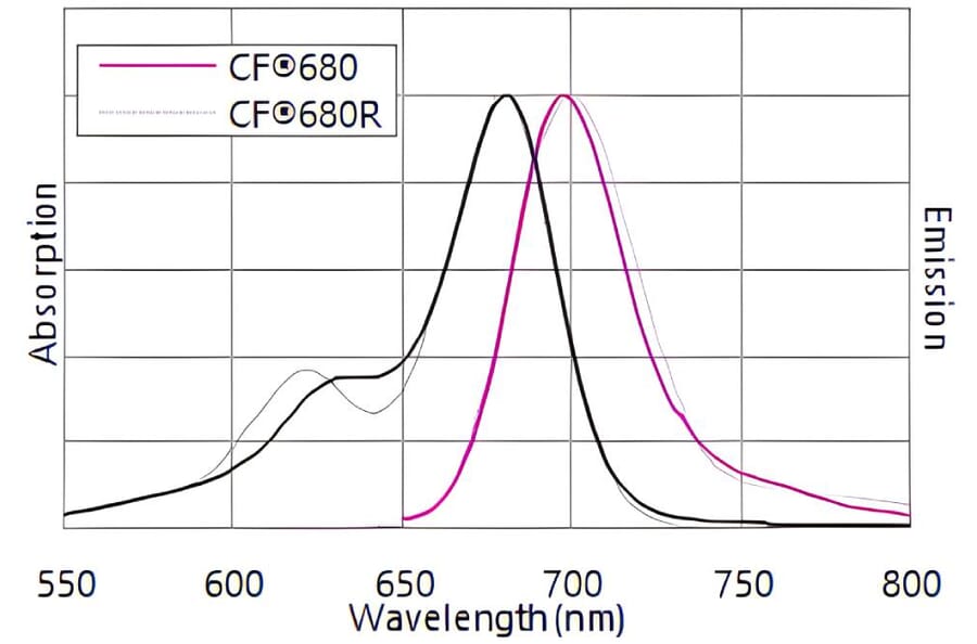
CF®680
Product Form
Liquid
Concentration
1 mg/mL
Formulation
Supplied in Phosphate Buffered Saline with 2 mg/ml BSA and 0.05% Sodium Azide.
Storage
Shipped at 4°C. Upon delivery aliquot and store at -20°C. Avoid freeze/thaw cycles. This product is also photosensitive and should be protected from light. Should this product contain a precipitate we recommend microcentrifugation before use. CF® Dyes are guaranteed for at least 6 months from data of receipt when stored correctly.
General Notes
Looking for a specific protein conjugate to simplify your workflow? We offer a library of over 2,000 targets conjugated to your choice of CF® dye. To enquire about a custom product, contact us directly.
Disclaimer
This product is for research use only. It is not intended for diagnostic or therapeutic use.
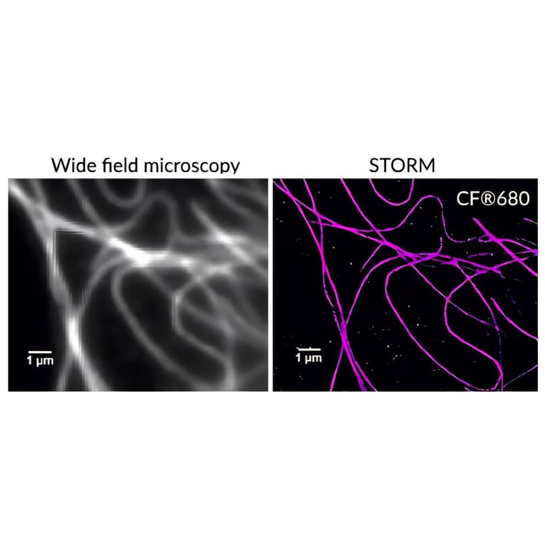
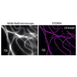
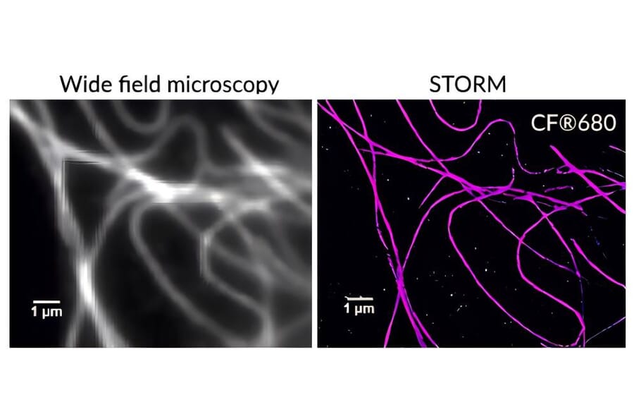
Comparison of microtubule imaging using conventional wide-field microscopy and STORM using a CF®680 dye conjugate. Images courtesy of Sam Kenny and Professor Ke Xu, College of Chemistry, University of California, Berkeley.
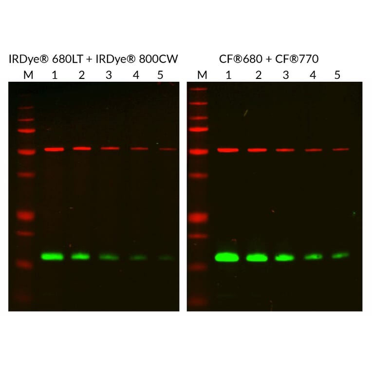
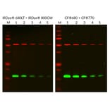
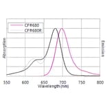
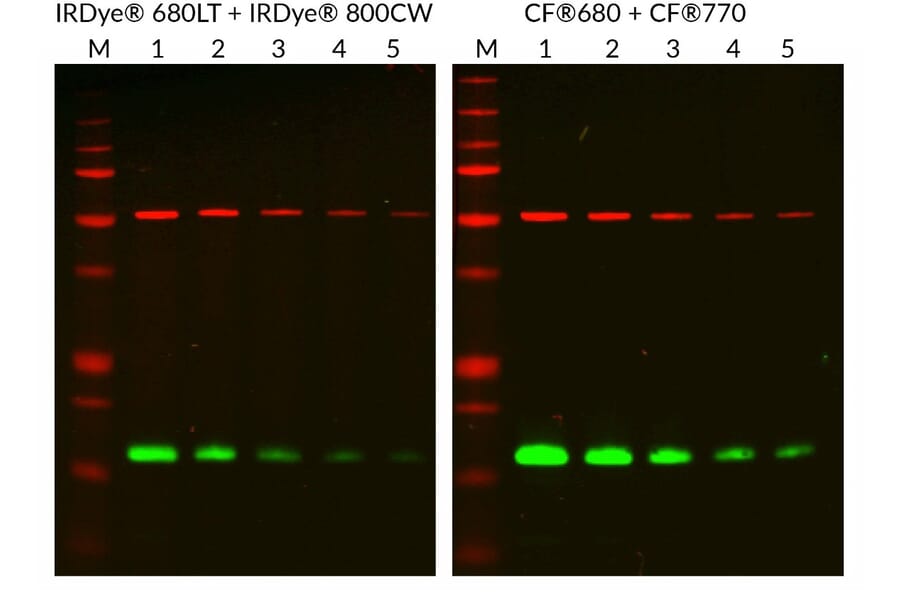
CF®680 provides brighter signal than IRDye® 680LT for near-infrared western blot detection. Two-fold dilutions of HeLa cell lysate (2 µg to 0.125 µg, lanes 1–5) were resolved and transferred to PVDF membranes. Mouse anti-tubulin and rabbit anti-COX IV primary antibodies were detected using IRDye® 680LT or CF®680 anti-mouse secondary antibodies (red) and IRDye® 800CW or CF®770 anti-rabbit secondary antibodies (green). Membranes were imaged on a LI-COR® Odyssey® NIR imaging system. Band quantification revealed approximately 50% higher signal intensity with CF® dyes compared to IRDye®. M indicates the molecular weight marker.
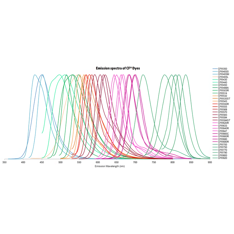
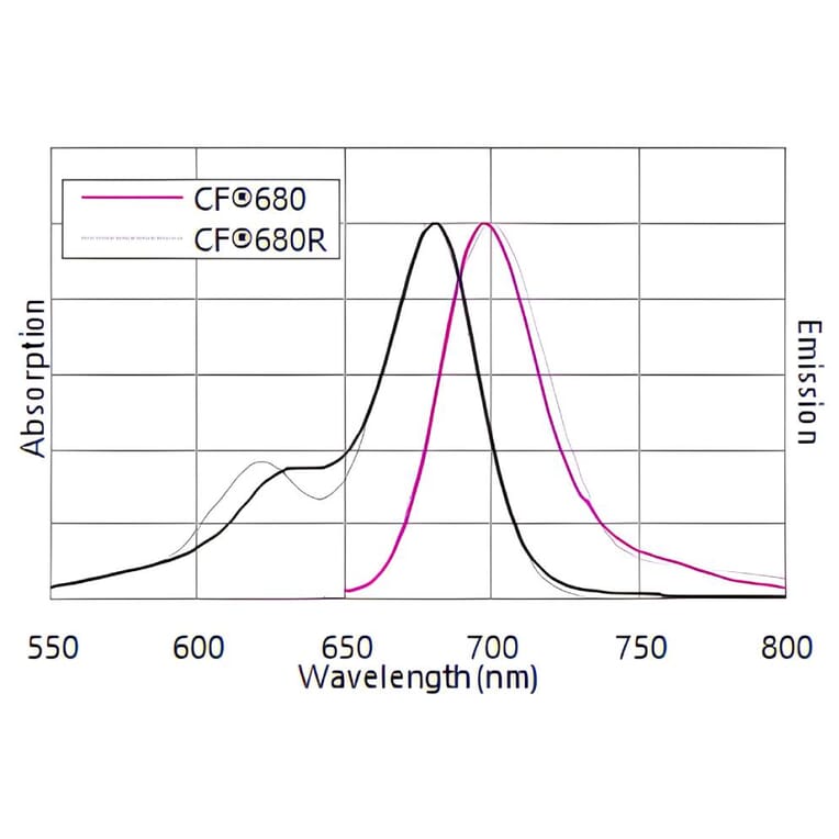
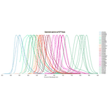
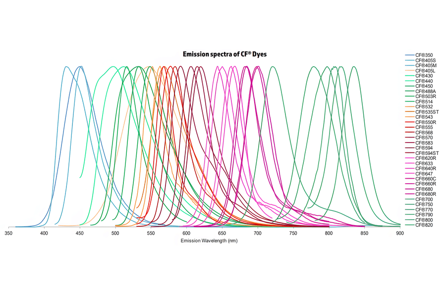
Normalized emission spectra of the CF® dye family spanning the visible to near-infrared range are shown, illustrating the spectral diversity and overlap between dyes. Curves represent relative fluorescence intensity as a function of emission wavelength (nm), with peak positions corresponding to each dye’s characteristic emission maximum. This reference highlights the broad coverage of CF® dyes for multicolor fluorescence applications and aids in selecting compatible dye combinations for imaging, flow cytometry, and other fluorescence-based assays.
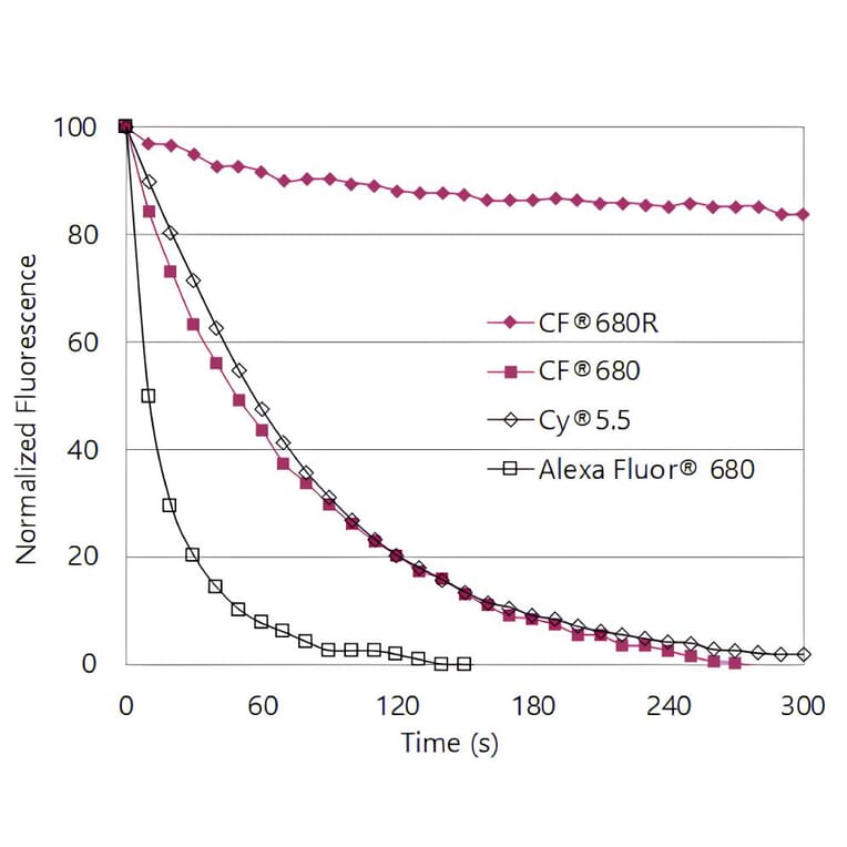
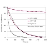
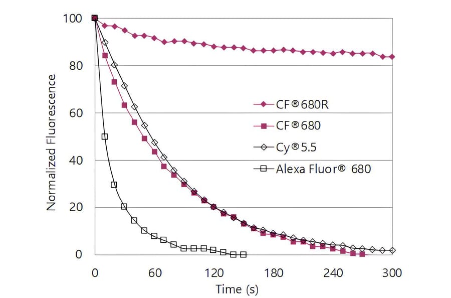
CF®680R is highly photostable. Jurkat cells were stained with mouse anti-CD3 antibody and goat anti-mouse conjugates of the dyes shown. Cells were exposed to continuous mercury arc lamp excitation with a Cy®5 filter set. Images were captured every 10 seconds for 5 minutes; mean fluorescence was normalized to time 0.
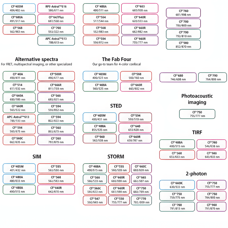
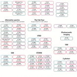
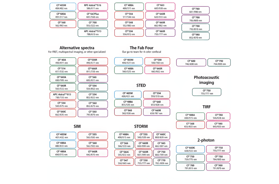
This chart summarizes commonly used CF® dyes grouped by their suitability for specific imaging modalities, including alternative spectra applications, four-color confocal imaging, near-infrared western blotting, photoacoustic imaging, STED, SIM, STORM, TIRF, and two-photon microscopy. Each dye is shown with its characteristic excitation and emission wavelengths (nm), providing a practical reference for selecting spectrally compatible dyes and optimizing multicolor experimental design across a range of fluorescence techniques.
Publishing research using Goat F(ab')2 Anti-Alpaca IgG, VHH domain + Llama IgG, VHH domain Antibody (CF®680) - Highly Cross-Adsorbed (A344044)? Please let us know so that we can list the citation on this page.