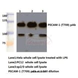
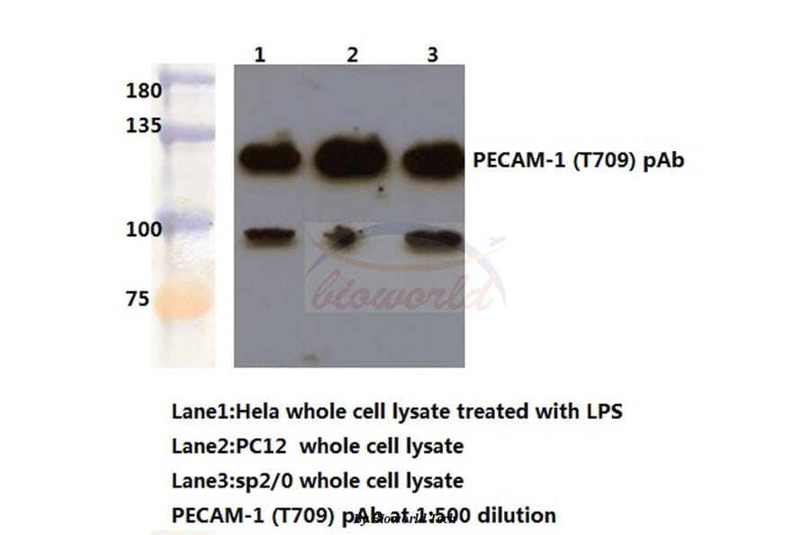
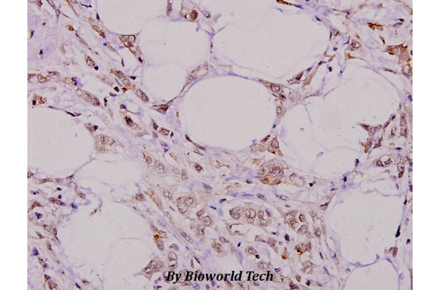
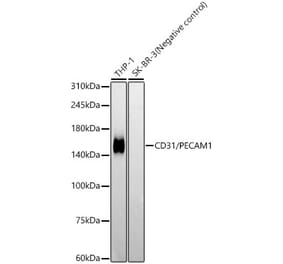
CD31/PECAM-1 (T709) pAb detects endogenous levels of CD31 protein.
Applications
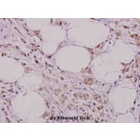
WB, IHC, IF
Reactivity
Human, Mouse, Rat
Immunogen
Synthetic peptide, corresponding to amino acids 670-720 of Human CD31.
Host
Rabbit
Clonality
Polyclonal
Conjugate
Unconjugated
Molecular Weight
~ 130 kDa
Purity
The antibody was affinity-purified from rabbit antiserum by affinity-chromatography using epitope-specific immunogen and the purity is > 95% (by SDS-PAGE).
Product Form
1 mg/ml in Phosphate buffered saline (PBS) with 0.05% sodium azide, approx. pH 7.2.


![Flow Cytometry - Anti-CD31 Antibody [MEM-05] (A86230)](https://cdn.antibodies.com/image/catalog/86/A86230_1.jpg?profile=product_alternative)
![Immunohistochemistry - Anti-CD31 Antibody [JC/70A] (A253621) - Antibodies.com](https://cdn.antibodies.com/image/catalog/249/A249604_1.jpg?profile=product_alternative)
![Immunohistochemistry - Anti-CD31 Antibody [JC/70A] - BSA and Azide free (A249604) - Antibodies.com](https://cdn.antibodies.com/image/catalog/252/A252784_1.jpg?profile=product_alternative)
![Immunohistochemistry - Anti-CD31 Antibody [C31.7] (A252781) - Antibodies.com](https://cdn.antibodies.com/image/catalog/249/A249599_1.jpg?profile=product_alternative)
![Immunohistochemistry - Anti-CD31 Antibody [PECAM1/3527] - BSA and Azide free (A252776) - Antibodies.com](https://cdn.antibodies.com/image/catalog/252/A252776_1.jpg?profile=product_alternative)
![Immunohistochemistry - Anti-CD31 Antibody [C31.7] - BSA and Azide free (A249599) - Antibodies.com](https://cdn.antibodies.com/image/catalog/252/A252779_1.jpg?profile=product_alternative)
![Immunohistochemistry - Anti-CD31 Antibody [ER-MP12] (A280366) - Antibodies.com](https://cdn.antibodies.com/image/catalog/280/A280366_4.jpg?profile=product_alternative)
![Immunohistochemistry - Anti-CD31 Antibody [PECAM1/3527] (A249596) - Antibodies.com](https://cdn.antibodies.com/image/catalog/249/A249596_1.jpg?profile=product_alternative)
![Immunohistochemistry - Anti-CD31 Antibody [PECAM1/3534] - BSA and Azide free (A278328) - Antibodies.com](https://cdn.antibodies.com/image/catalog/278/A278328_1.jpg?profile=product_alternative)
![Immunohistochemistry - Anti-CD31 Antibody [C31.3] (A252786) - Antibodies.com](https://cdn.antibodies.com/image/catalog/249/A249601_1.jpg?profile=product_alternative)
![Immunohistochemistry - Anti-CD31 Antibody [PECAM1/3534] (A277740) - Antibodies.com](https://cdn.antibodies.com/image/catalog/277/A277740_1.jpg?profile=product_alternative)