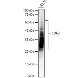
CD63 (K156) pAb detects endogenous levels of CD63 protein.
Applications
WB, IHC
Reactivity
Human, Mouse, Rat
Immunogen
Synthetic peptide, corresponding to amino acids 111-160 of Human CD63.
Host
Rabbit
Clonality
Polyclonal
Conjugate
Unconjugated
Molecular Weight
~ 17, 23, 26kDa
Purity
The antibody was affinity-purified from rabbit antiserum by affinity-chromatography using epitope-specific immunogen and the purity is > 95% (by SDS-PAGE).
Product Form
1 mg/ml in Phosphate buffered saline (PBS) with 0.05% sodium azide, approx. pH 7.2.
Synonyms
CD63 antigen, Granulophysin, LAMP-3, Limp1, Lysosomal-associated membrane protein 3, Lysosome integral membrane protein 1, Melanoma-associated antigen ME491, MLA1, OMA81H, TSPAN30


![ICC - Anti-CD63 Antibody [MEM-259] (A85976)](https://cdn.antibodies.com/image/catalog/85/A85976_1.jpg?profile=product_alternative)


![Immunohistochemistry - Anti-CD63 Antibody [MX-49.129.5] (A253930) - Antibodies.com](https://cdn.antibodies.com/image/catalog/250/A250748_1.jpg?profile=product_alternative)
![Immunohistochemistry - Anti-CD63 Antibody [NKI/C3] - BSA and Azide free (A250746) - Antibodies.com](https://cdn.antibodies.com/image/catalog/253/A253926_1.jpg?profile=product_alternative)
![Immunohistochemistry - Anti-CD63 Antibody [MX-49.129.5] - BSA and Azide free (A250748) - Antibodies.com](https://cdn.antibodies.com/image/catalog/253/A253928_1.jpg?profile=product_alternative)
![Immunohistochemistry - Anti-CD63 Antibody [NKI/C3] (A253928) - Antibodies.com](https://cdn.antibodies.com/image/catalog/250/A250746_1.jpg?profile=product_alternative)

![Immunohistochemistry - Anti-CD63 Antibody [LAMP3/2789] - BSA and Azide free (A253934) - Antibodies.com](https://cdn.antibodies.com/image/catalog/253/A253934_1.jpg?profile=product_alternative)
![Immunohistochemistry - Anti-CD63 Antibody [LAMP3/2789] (A250754) - Antibodies.com](https://cdn.antibodies.com/image/catalog/250/A250754_1.jpg?profile=product_alternative)
![Immunohistochemistry - Anti-CD63 Antibody [rMX-49.129.5] (A253926) - Antibodies.com](https://cdn.antibodies.com/image/catalog/250/A250752_1.jpg?profile=product_alternative)