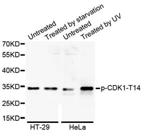
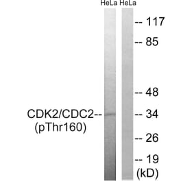
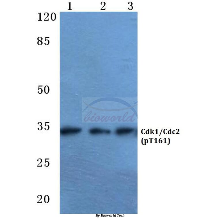
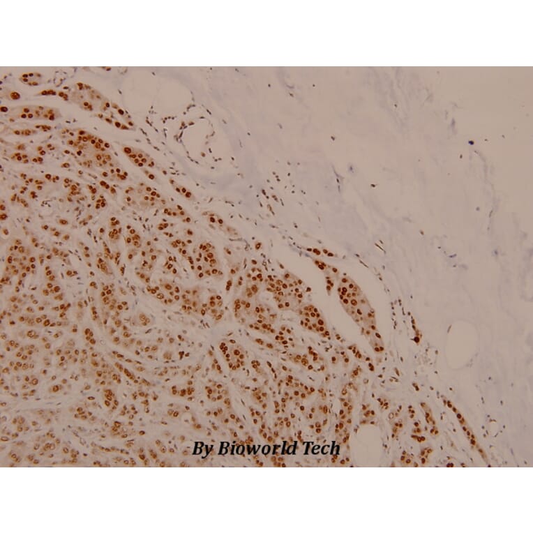
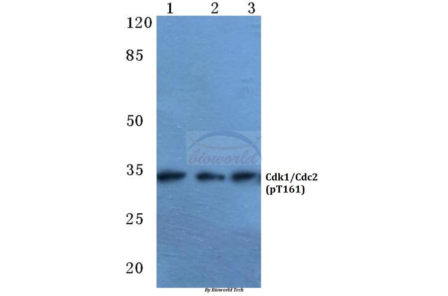
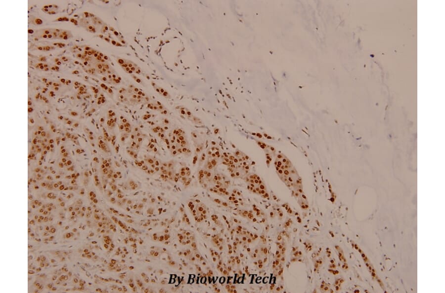
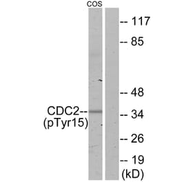
Rabbit polyclonal antibody to Cdk1/Cdc2 (phospho-T161)
Specificity
p-Cdk1/Cdc2 (T161) pAb detects endogenous levels of p-Cdk1/Cdc2 protein only when phosphorylated at Thr161.
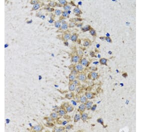
Applications
WB, IHC
Reactivity
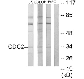
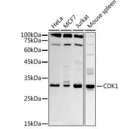
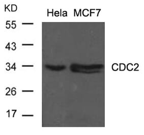
Human, Mouse, Rat
Immunogen
Synthetic phosphopeptide derived from human Cdk1/Cdc2 around the phosphorylation site of Serine 795.
Host
Rabbit
Clonality
Polyclonal
Conjugate
Unconjugated
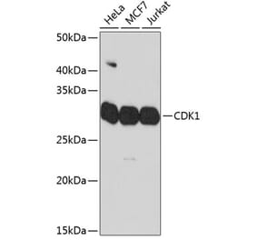
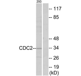
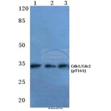
Molecular Weight
~ 34 kDa
Purity
The antibody was affinity-purified from rabbit antiserum by affinity-chromatography using epitope-specific immunogen and the purity is > 95% (by SDS-PAGE).
Product Form
1 mg/ml in Phosphate buffered saline (PBS) with 15 mM sodium azide, approx. pH 7.2.
Synonyms
CDC2, CDC28A, CDKN1, Cell division control protein 2 homolog, Cell division protein kinase 1, Cyclin-dependent kinase 1, p34 protein kinase, P34CDC2


![IHC - Anti-CDK1 Antibody [POH-1] (A86650)](https://cdn.antibodies.com/image/catalog/86/A86650_1.jpg?profile=product_alternative)
![Immunohistochemistry - Anti-CDK1 Antibody [A17.1.1] (A253992) - Antibodies.com](https://cdn.antibodies.com/image/catalog/250/A250808_1.jpg?profile=product_alternative)
![Immunohistochemistry - Anti-CDK1 Antibody [A17.1.1] - BSA and Azide free (A250808) - Antibodies.com](https://cdn.antibodies.com/image/catalog/253/A253988_1.jpg?profile=product_alternative)