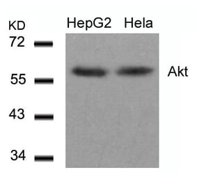
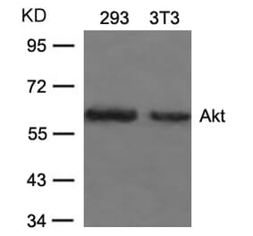
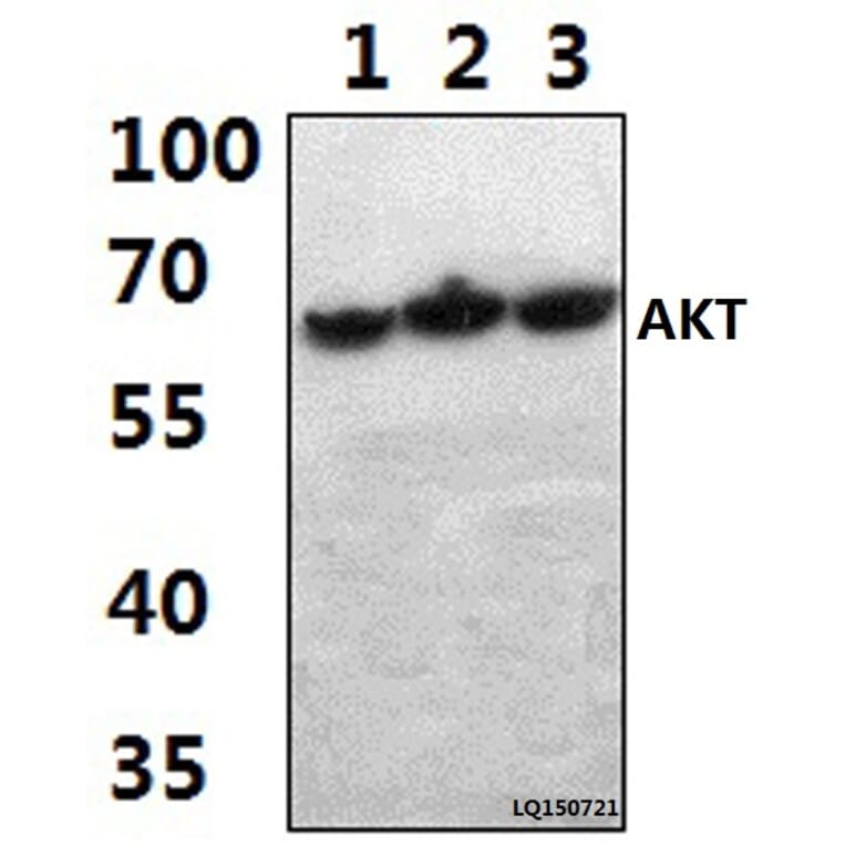
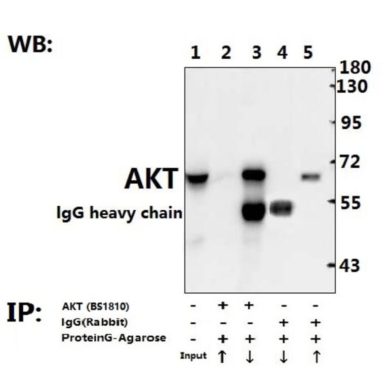
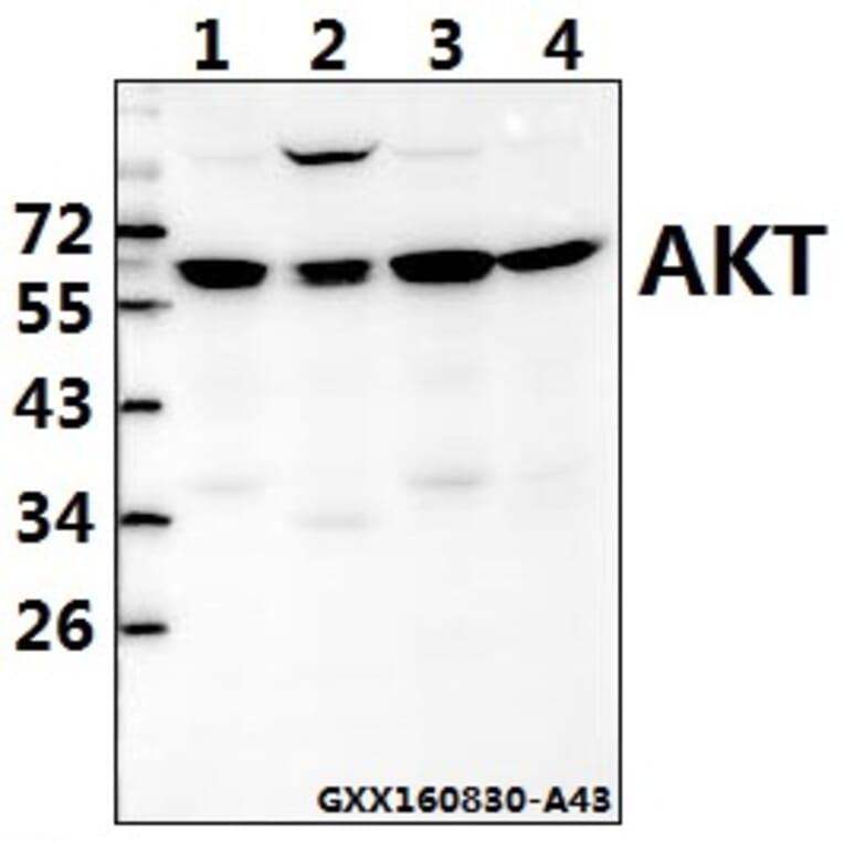
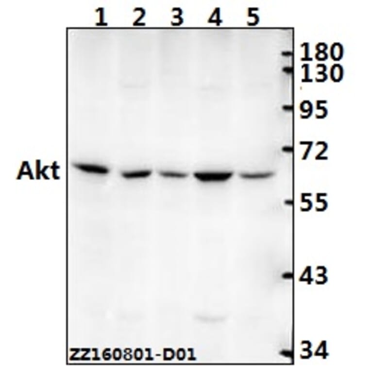
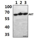
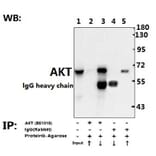
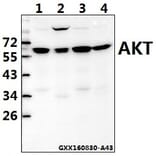
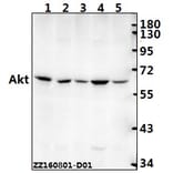
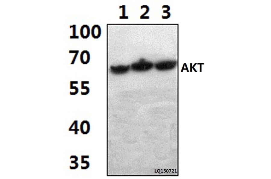
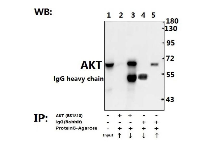
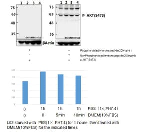
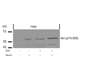
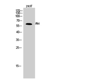
AKT (A444) pAb detects endogenous levels of total AKT protein.
Applications
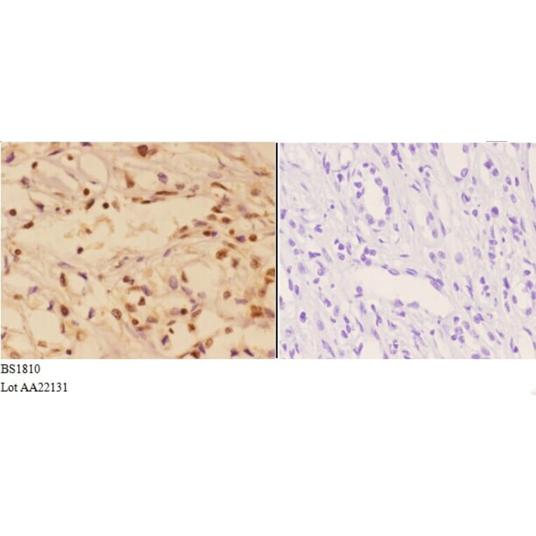
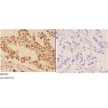
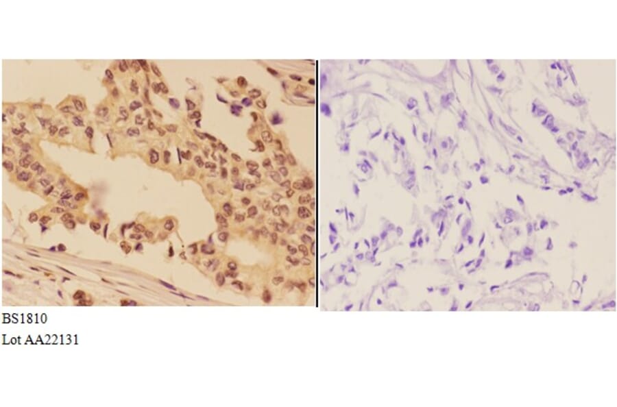
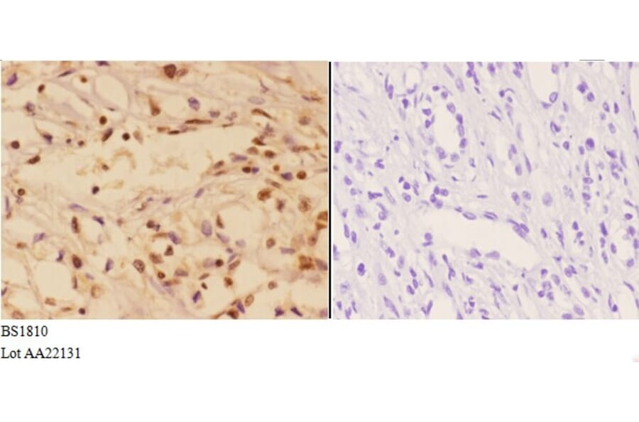
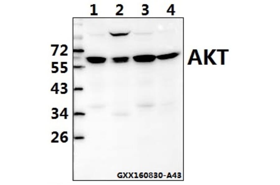
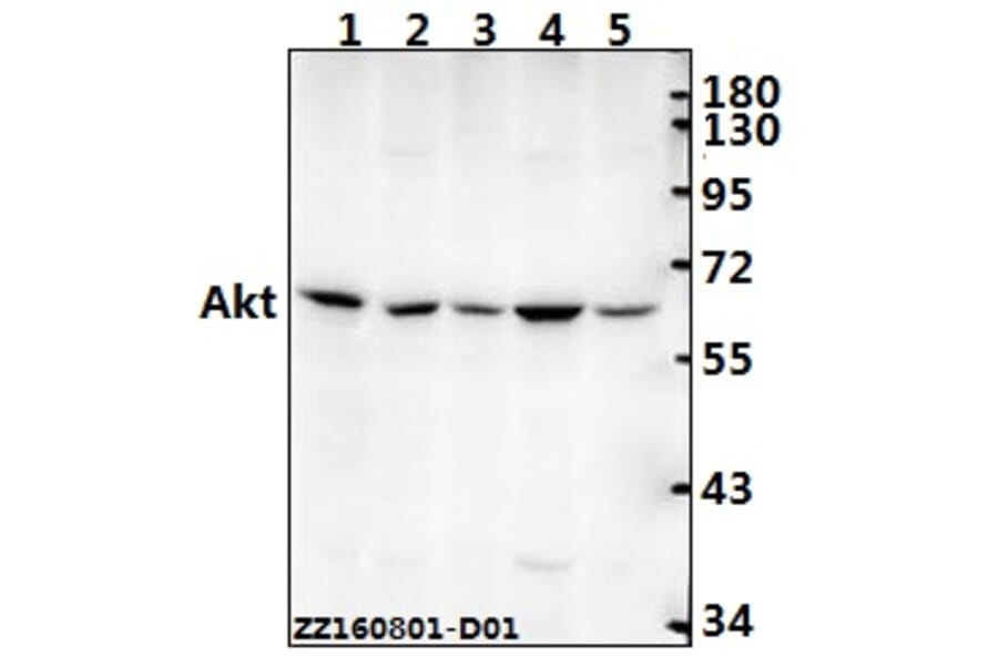
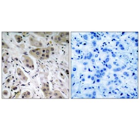
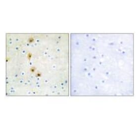
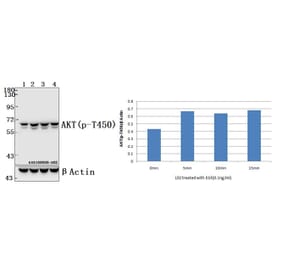
WB, IHC
Reactivity
Human, Mouse, Rat
Immunogen
Synthetic peptide, corresponding to amino acids 420-470 of Human AKT1.
Host
Rabbit
Clonality
Polyclonal
Conjugate
Unconjugated
Molecular Weight
~ 60 kDa
Purity
The antibody was affinity-purified from rabbit antiserum by affinity-chromatography using epitope-specific immunogen and the purity is > 95% (by SDS-PAGE).
Product Form
1 mg/ml in Phosphate buffered saline (PBS) with 0.05% sodium azide, approx. pH 7.2.


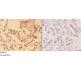
![Immunohistochemistry - Anti-AKT Antibody [RM316] (A121372) - Antibodies.com](https://cdn.antibodies.com/image/catalog/121/A121394_1.png?profile=product_alternative)