

Unconjugated
Senescent neural progenitor cells have been identified in brain lesions of people with progressive multiple sclerosis (PMS). However, their role in disease pathobiology and contribution to the lesion environment remains unclear. By establishing directly induced neural stem/progenitor cell (iNSC) lines from PMS patient fibroblasts, we studied their senescent phenotype in vitro. Senescence was strongly associated with inflammatory signaling, hypermetabolism, and the senescence-associated secretory phenotype (SASP). PMS-derived iNSCs displayed increased glucose-dependent fatty acid and cholesterol synthesis, which resulted in the accumulation of lipid droplets. A 3-hydroxy-3-methylglutaryl (HMG)-coenzyme A (CoA) reductase (HMGCR)-mediated lipogenic state was found to induce a SASP in PMS iNSCs via cholesterol-dependent transcription factors. SASP from PMS iNSC lines induced neurotoxicity in mature neurons, and treatment with the HMGCR inhibitor simvastatin altered the PMS iNSC SASP, promoting cytoprotective qualities and reducing neurotoxicity. Our findings suggest a disease-associated, cholesterol-related, hypermetabolic phenotype of PMS iNSCs that leads to neurotoxic signaling and is rescuable pharmacologically.
Objectives: Sjögren's disease (SjD) is a systemic autoimmune disorder characterized by dysfunction of exocrine glands, particularly the salivary and lacrimal glands, with no clear etiology or effective therapy. This study explores the complex interplay of varied cell types in the salivary glands and their role in the pathology of Sjögren's disease.
Methods: Utilizing single-cell and spatial transcriptomics alongside spatial immunophenotyping to analyze human minor salivary glands, we developed a comprehensive understanding of the cellular landscape of non-SjD salivary glands and how that landscape changes in SjD patients. In vitro cellular assays and novel patient-derived primary epithelial cells were co-cultured with autologous T cells to confirm effector states and the delivery and effect of disease-associated granzymes.
Results: We identified previously unrecognized heterogeneity among acinar cells, including a PRR4?CST3?WFDC2? seromucous acinar population that is selectively lost in Sjögren's disease. Expression and organizational changes were linked to clinical features: (i) T cells in the glands of SSA?, high-focus score patients showed increased transcriptional signatures of activation, antigen presentation, and apoptosis resistance compared with patients with mild or moderate disease, and (ii) patients with low immune infiltration exhibited distinct epithelial organization. Notably, GZMK?CD8? T cells, which accumulate with disease severity, displayed a cytotoxic transcriptional program, degranulated upon stimulation ex vivo, and localized spatially with immune-engaged epithelial cells. Functional assays demonstrated that GZMK activates interferon signaling in vitro, and autologous co-cultures of patient-derived T cells and epithelial cells validated these findings.
Conclusions: Using single-cell and spatial transcriptomics and proteomics, this study identifies a selective loss of PRR4?CST3?WFDC2? seromucous acinar cells and a rise in GZMK?CD8? T cells in Sjögren's disease, revealing distinct immune-mediated epithelial remodeling and interferon-driven dysfunction across diverse clinical presentations. These findings uncover a novel sub-cytolytic effector mechanism by which GZMK?CD8? T cells impair mitochondrial integrity and activate innate immune signaling, linking epithelial injury to type I interferon responses and offering new therapeutic targets.
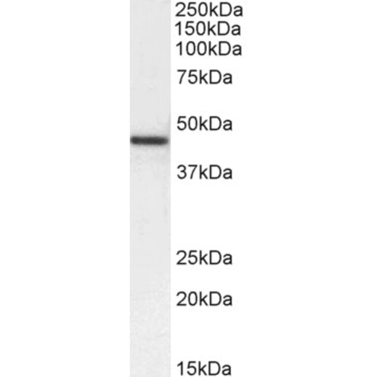
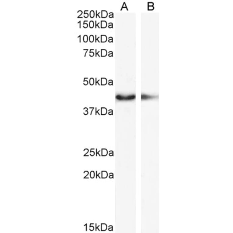




![Immunohistochemistry - Anti-alpha Smooth Muscle Actin Antibody [1A4] (A253077) - Antibodies.com](https://cdn.antibodies.com/image/catalog/249/A249894_1.jpg?profile=product_alternative)
![Immunohistochemistry - Anti-alpha Smooth Muscle Actin Antibody [1A4] - BSA and Azide free (A249894) - Antibodies.com](https://cdn.antibodies.com/image/catalog/253/A253074_1.jpg?profile=product_alternative)
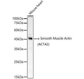

![Immunohistochemistry - Anti-alpha Smooth Muscle Actin Antibody [SPM332] (A253076) - Antibodies.com](https://cdn.antibodies.com/image/catalog/249/A249895_1.jpg?profile=product_alternative)
![Immunohistochemistry - Anti-alpha Smooth Muscle Actin Antibody [SPM332] - BSA and Azide free (A249895) - Antibodies.com](https://cdn.antibodies.com/image/catalog/253/A253075_1.jpg?profile=product_alternative)
![Immunofluorescence - Anti-alpha Smooth Muscle Actin Antibody [HHF35] (A115508) - Antibodies.com](https://cdn.antibodies.com/image/catalog/115/A115508_1.jpg?profile=product_alternative)
![Immunofluorescence - Anti-alpha Smooth Muscle Actin Antibody [1A4] (A115506) - Antibodies.com](https://cdn.antibodies.com/image/catalog/115/A115506_1.jpg?profile=product_alternative)
![Immunohistochemistry - Anti-alpha Smooth Muscle Actin Antibody [ACTA2/791] (A253077) - Antibodies.com](https://cdn.antibodies.com/image/catalog/249/A249896_1.jpg?profile=product_alternative)
![Immunohistochemistry - Anti-alpha Smooth Muscle Actin Antibody [1A4 + ACTA2/791] (A252041) - Antibodies.com](https://cdn.antibodies.com/image/catalog/249/A249897_1.jpg?profile=product_alternative)
![Immunohistochemistry - Anti-alpha Smooth Muscle Actin Antibody [ACTA2/791] - BSA and Azide free (A249896) - Antibodies.com](https://cdn.antibodies.com/image/catalog/253/A253076_1.jpg?profile=product_alternative)
![Immunohistochemistry - Anti-alpha Smooth Muscle Actin Antibody [1A4 + ACTA2/791] - BSA and Azide free (A249897) - Antibodies.com](https://cdn.antibodies.com/image/catalog/253/A253077_1.jpg?profile=product_alternative)