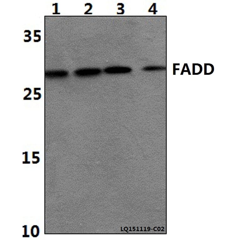
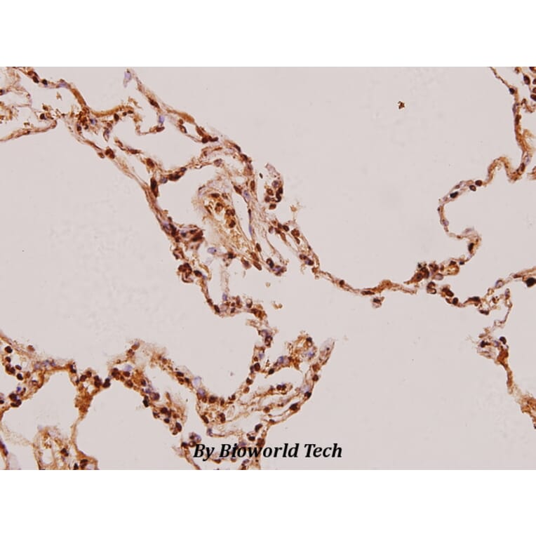
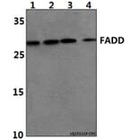
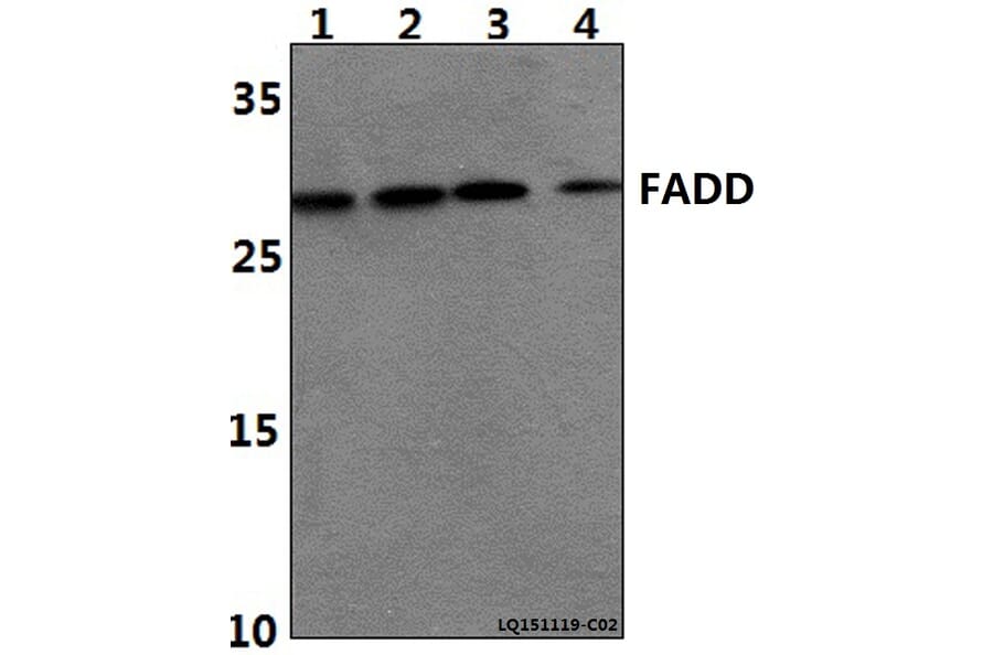
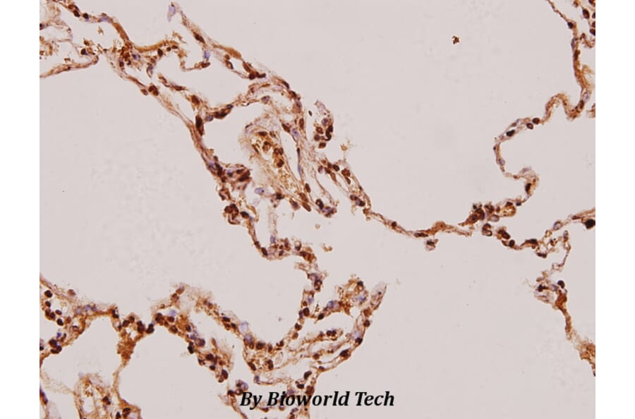
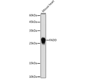
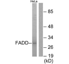
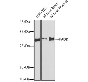
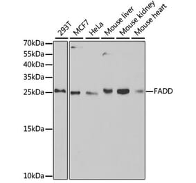
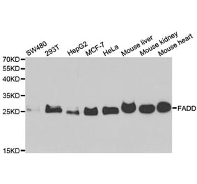
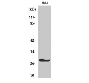
Unconjugated
Actinomycin D (Act D), a well known of clinical antitumor drug, has been used for the treatment of some highly malignant tumors, however, the clinical application was limited by its extreme cytotoxicity. In the present study, we reported that methylated actinomycin D (mAct D), a novel actinomycin D analog isolated from Streptomyces sp. KLBMP 2541 in our previous study, could not only exert stronger inhibitory effects on several human cancer cells than Act D in dose- and time-dependent manner at ng concentrations, especially on HepG2 cells, but also lower cytotoxicity in normal cells (HL-7702). Base on these results, HepG2 cells were treated for further study to illustrate the potential mechanism of mAct D. The results of nuclei morphology examination, DNA fragmentation detection, sub-G1 analysis, annexin V-FITC/PI staining and activation of caspase-3 indicated mAct D significantly induced HepG2 cells apoptosis. Semiquantitative RT-PCR and Western blot analysis revealed that mAct D induced apoptosis in HepG2 cells through mitochondria-dependent pathway by increasing levels of caspase-9, Bax, Bak while decreasing levels of Bcl-2, Bid, and Fas-dependent pathway by increasing levels of Fas, FasL, FADD, and caspase-8. Subsequently, pretreatment with specific inhibitor of caspase-8 Z-LEHD-FMK and caspase-9 Z-LEHD-FMK significantly attenuated caspase-3 activity, the cleavage of caspase-3 and PARP, meanwhile increased the cell viability. In addition, p53 and mitochondrial transcription factor A (mtTFA) were also upregulated. Taken together, ng concentrations mAct D induces the apoptosis of HepG2 through Fas- and mitochondria-mediated pathway and presents a potential novel alternative agent for the treatment of human hepatic carcinoma.
In the present study, the antiproliferative effect of dioscin on human gastric carcinoma SGC-7901 cells was confirmed by 3-(4, 5-dimethylthiahiazo-zyl)-2, 5-dip-henytetrazolium bromide and flow cytometry assays. Through acridine orange-ethidium bromide double fluorescent staining, apoptotic morphology of the cells was observed. Radioimmunoassays showed that the tumor necrosis factor (TNF)-α concentration in cells treated with dioscin significantly increased compared with untreated cells. Several proteins and mRNA related to the mitochondrial and death receptor pathways were investigated. We found that the expression of Bid, bcl-2 and bcl-xl was markedly downregulated, and the expression of Bak and Bax was upregulated. In addition, cytochrome c was released from the mitochondria into the cytosol, which indicates activation of the mitrochondrial pathway by dioscin. Furthermore, upregulation of Fas, FasL (Fas ligand), TNF-α, TNF receptor-1, TNF receptor-associated factor 1 and Fas-associated protein with death domain demonstrated involvement of the death receptor pathway. Increased mRNA expression of p53 was also found in dioscin-treated SGC-7901 cells, and the activation of caspase-3 and -8 was also observed. Consequently, this study clarifies the mechanism underlying the anticancer effect of dioscin, and also indicates that dioscin may be a potential drug treatment for human gastric cancer.