

Unconjugated
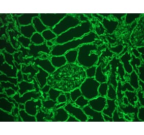
Collagen type-IV (Col-IV) is the principal basement membrane structural component of the blood-gas barrier (BGB) to which the strength, i.e., the structural integrity, of the profoundly thin tissue barrier of the lung has been attributed. In this study, Col-IV was immunolocalized and the spatial organization, and relative volume density in the exchange tissues (ETs) of the lung of an adult common quail (Coturnix coturnix) was determined. Formalin-fixed paraffin-embedded lung sections were immunolabeled with anti-collagen IV monoclonal primary antibody and detected using Alexa Fluor 488-conjugated secondary antibody, prior to confocal laser scanning microscopy. Three-dimensional reconstructions were prepared from the acquired image datasets to allow visualization of the spatial arrangement of Col-IV in the ETs. The volume densities of Col-IV in the BGB, the epithelial-epithelial cell connections, and the blood capillary-blood capillary connections were determined by the stereological method of point-counting. Three-dimensional reconstructions showed heterogeneous, vast distribution of Col-IV within the BGB. In the parabronchial ETs, Col-IV continues centripetally, i.e., inwards, to connect to the atrial smooth muscles. The structural feature shows existence of a functional continuum that may strengthen the parabronchus, contributing to the rigidity of the air- and the blood capillaries. The high-volume density of Col-IV in the ETs (~20%) of the lungs of the investigated adult common quail indicates its significance as a structural component. The abundance and the organization of Col-IV in the ETs underpins its importance in stabilizing the lung histo-architecture, particularly the exceptionally thin BGB. The findings provide insights that may inform on optimal biomimetic designs of artificial gas-exchange membranes.
A minimal diffusion barrier is key to the pulmonary gas exchange. In alveolar capillary dysplasia (ACD), a rare genetically driven disease of early infancy, this crucial fibrovascular interface is compromised while the underlying pathophysiology is insufficiently understood. Recent in-depth analyses of vascular alterations in adult lung disease encouraged researchers to extend these studies to ACD and compare the changes of the microvasculature. Lung tissue samples of children with ACD (n = 12), adults with non-specific interstitial pneumonia (n = 12), and controls (n = 20) were studied using transmission electron microscopy, single-gene sequencing, immunostaining, exome sequencing, and broad transcriptome profiling. In ACD, pulmonary capillary basement membranes were hypertrophied, thickened, and multilamellated. Transcriptome profiling revealed increased CDH5, COL4A1, COL15A1, PTK2B, and FN1 and decreased VIT expression, confirmed by immunohistochemistry. In contrast, non-specific interstitial pneumonia samples showed a regular basement membrane architecture with preserved VIT expression but also increased COL15A1+ vessels. This study provides insight into the ultrastructure and pathophysiology of ACD. The lack of normally developed lung capillaries appeared to cause a replacement by COL15A1+ vessels, a mechanism recently described in interstitial lung disease. The VIT loss and FN1 overexpression might contribute to the unique appearance of basement membranes in ACD. Future studies are needed to explore the therapeutic potential of down-regulating the expression of FN1 and balancing VIT deficiency.
![Immunohistochemistry - Anti-Collagen IV Antibody [M3F7] (A248246) - Antibodies.com](https://cdn.antibodies.com/image/catalog/248/A248246_1.jpg?profile=product_top)
![Immunohistochemistry - Anti-Collagen IV Antibody [M3F7] (A252777) - Antibodies.com](https://cdn.antibodies.com/image/catalog/248/A248246_2.jpg?profile=product_top)
![Immunofluorescence - Anti-Collagen IV Antibody [M3F7] (A248246) - Antibodies.com Citation](https://cdn.antibodies.com/image/catalog/248/A248246_100235.png?profile=product_top)
![Immunofluorescence - Anti-Collagen IV Antibody [M3F7] (A248246) - Antibodies.com Citation](https://cdn.antibodies.com/image/catalog/248/A248246_100237.png?profile=product_top)
![Immunofluorescence - Anti-Collagen IV Antibody [M3F7] (A248246) - Antibodies.com Citation](https://cdn.antibodies.com/image/catalog/248/A248246_100239.png?profile=product_top)
![Immunofluorescence - Anti-Collagen IV Antibody [M3F7] (A248246) - Antibodies.com Citation](https://cdn.antibodies.com/image/catalog/248/A248246_100241.png?profile=product_top)
![Immunohistochemistry - Anti-Collagen IV Antibody [M3F7] (A248246) - Antibodies.com](https://cdn.antibodies.com/image/catalog/248/A248246_1.jpg?profile=product_top_thumb)
![Immunohistochemistry - Anti-Collagen IV Antibody [M3F7] (A252777) - Antibodies.com](https://cdn.antibodies.com/image/catalog/248/A248246_2.jpg?profile=product_top_thumb)
![Immunofluorescence - Anti-Collagen IV Antibody [M3F7] (A248246) - Antibodies.com Citation](https://cdn.antibodies.com/image/catalog/248/A248246_100235.png?profile=product_top_thumb)
![Immunofluorescence - Anti-Collagen IV Antibody [M3F7] (A248246) - Antibodies.com Citation](https://cdn.antibodies.com/image/catalog/248/A248246_100237.png?profile=product_top_thumb)
![Immunofluorescence - Anti-Collagen IV Antibody [M3F7] (A248246) - Antibodies.com Citation](https://cdn.antibodies.com/image/catalog/248/A248246_100239.png?profile=product_top_thumb)
![Immunofluorescence - Anti-Collagen IV Antibody [M3F7] (A248246) - Antibodies.com Citation](https://cdn.antibodies.com/image/catalog/248/A248246_100241.png?profile=product_top_thumb)
![Immunohistochemistry - Anti-Collagen IV Antibody [M3F7] (A248246) - Antibodies.com](https://cdn.antibodies.com/image/catalog/248/A248246_1.jpg?profile=product_image)
![Immunohistochemistry - Anti-Collagen IV Antibody [M3F7] (A252777) - Antibodies.com](https://cdn.antibodies.com/image/catalog/248/A248246_2.jpg?profile=product_image)
![Immunofluorescence - Anti-Collagen IV Antibody [M3F7] (A248246) - Antibodies.com Citation](https://cdn.antibodies.com/image/catalog/248/A248246_100235.png?profile=product_image)
![Immunofluorescence - Anti-Collagen IV Antibody [M3F7] (A248246) - Antibodies.com Citation](https://cdn.antibodies.com/image/catalog/248/A248246_100237.png?profile=product_image)
![Immunofluorescence - Anti-Collagen IV Antibody [M3F7] (A248246) - Antibodies.com Citation](https://cdn.antibodies.com/image/catalog/248/A248246_100239.png?profile=product_image)
![Immunofluorescence - Anti-Collagen IV Antibody [M3F7] (A248246) - Antibodies.com Citation](https://cdn.antibodies.com/image/catalog/248/A248246_100241.png?profile=product_image)

![Immunohistochemistry - Anti-Collagen IV Antibody [M3F7] - BSA and Azide free (A251428) - Antibodies.com](https://cdn.antibodies.com/image/catalog/251/A251428_1.jpg?profile=product_alternative)

![Immunohistochemistry - Frozen - Anti-Collagen IV Antibody [1043] (A115551) - Antibodies.com](https://cdn.antibodies.com/image/catalog/115/A115551_1.png?profile=product_alternative)
![Immunohistochemistry - Anti-Collagen IV Antibody [IHC549] (A86892) - Antibodies.com](https://cdn.antibodies.com/image/catalog/86/A86892_1.jpg?profile=product_alternative)
![Immunohistochemistry - Anti-Collagen IV Antibody [rCOL4/4742] - BSA and Azide free (A251427) - Antibodies.com](https://cdn.antibodies.com/image/catalog/251/A251427_1.jpg?profile=product_alternative)
![Immunohistochemistry - Anti-Collagen IV Antibody [COL4/4241R] - BSA and Azide free (A251429) - Antibodies.com](https://cdn.antibodies.com/image/catalog/251/A251429_1.jpg?profile=product_alternative)
![Immunohistochemistry - Anti-Collagen IV Antibody [rCOL4/4742] (A248245) - Antibodies.com](https://cdn.antibodies.com/image/catalog/248/A248245_1.jpg?profile=product_alternative)