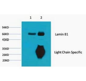
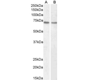
Synthetic peptide, corresponding to amino acids 50-100 of Human Lamin B1.
Host
Rabbit
Clonality
Polyclonal
Conjugate
Unconjugated
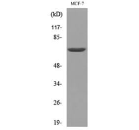
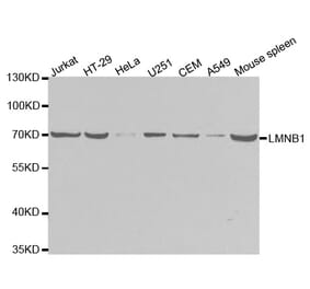
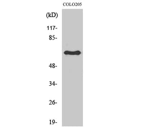
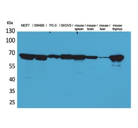
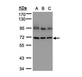
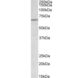
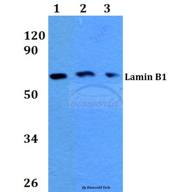
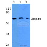
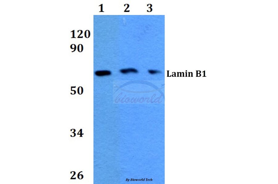
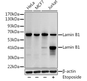
Molecular Weight
~ 68 kDa
Purity
The antibody was affinity-purified from rabbit antiserum by affinity-chromatography using epitope-specific immunogen and the purity is > 95% (by SDS-PAGE).
Product Form
1 mg/ml in Phosphate buffered saline (PBS) with 0.05% sodium azide, approx. pH 7.2.


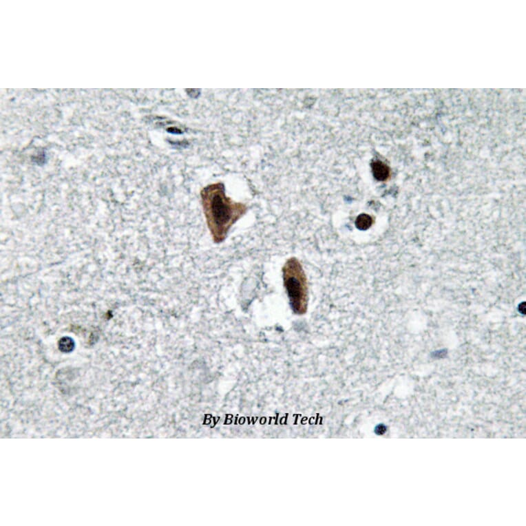
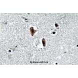
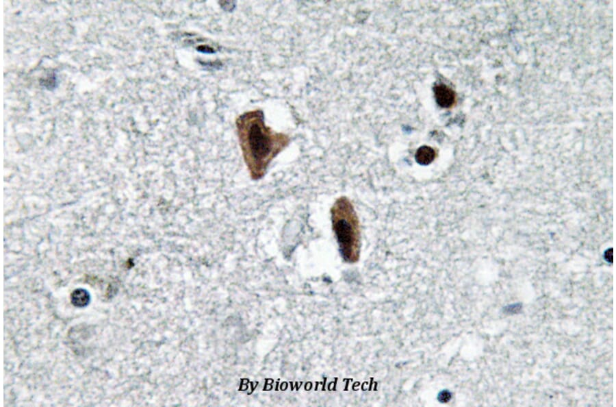
![Immunohistochemistry - Anti-Lamin B1 Antibody [119D5-F1] (A115608) - Antibodies.com](https://cdn.antibodies.com/image/catalog/115/A115608_1.jpg?profile=product_alternative)