Primary Antibodies
Secondary Antibodies
Proteins & Peptides
ELISA Kits
About Us
Contact Us
Sign In/Register
0
ISO 9001:2015 Certified
Live Customer Support
4.5/5 on Trustpilot
100% Quality Guarantee
Home
Primary Antibodies
mCherry Antibodies
Anti-mCherry Antibody (A121571)
Anti-mCherry Antibody (A121571)
Overview
Specifications
Images
Enlarge Image
Enlarge Image
Enlarge Image
Enlarge Image
$590
Product Datasheet
Goat polyclonal antibody to mCherry for WB, IF, IHC-P, IHC-Fr and IEM.
100% Guarantee
Price Match Guarantee
Product Size:
600µg
1.5mg
Quantity:
1
2
3
4
5
6
7
8
9
10
Add To Cart
Request a Quotation
Custom or Bulk Request
Shipping Information
Freight/Packing Charges:
$40
Dispatched from St. Louis, MO.
Lead Time: 5-8 business days.
Specifications
Name
Anti-mCherry Antibody
Description
Goat polyclonal antibody to mCherry.
Applications
WB
,
IF
,
IHC-P
,
IHC-Fr
, IEM
Dilutions
WB: 1:500-1:5,000, IF: 50-1:500, IHC-P: 1:50-1:500, IHC-Fr: 1:50-1:500
Reactivity
mCherry, tdTomato, Red Fluorescent Protein (dsRed)
Cross Reactivity
This antibody does not cross-react with green fluorescent protein.
Immunogen
Recombinant mCherry protein, expressed in and purified from E. coli.
Host
Goat
Clonality
Polyclonal
Isotype
IgG
Conjugate
Unconjugated
Purification
Affinity purification.
Concentration
3 mg/ml
Product Form
Liquid
Formulation
Supplied in Phosphate Buffered Saline with 20% Glycerol and 0.05% Sodium Azide.
Storage
Shipped at 4°C. Upon delivery aliquot and store at -20°C. Avoid freeze/thaw cycles.
Isotype Controls
Goat IgG (A121671)
Suitable Secondaries
Donkey Anti-Goat IgG H&L Antibody (AP) (A300679)
Donkey Anti-Goat IgG H&L Antibody (Biotin) (A300716)
Donkey Anti-Goat IgG H&L Antibody (FITC) (A300685)
Donkey Anti-Goat IgG H&L Antibody (HRP) (A300730)
See all Anti-Goat IgG Secondaries →
Disclaimer
This product is for research use only. It is not intended for diagnostic or therapeutic use.
Scientific Validation Data
Validation Data
(4)
Enlarge Image
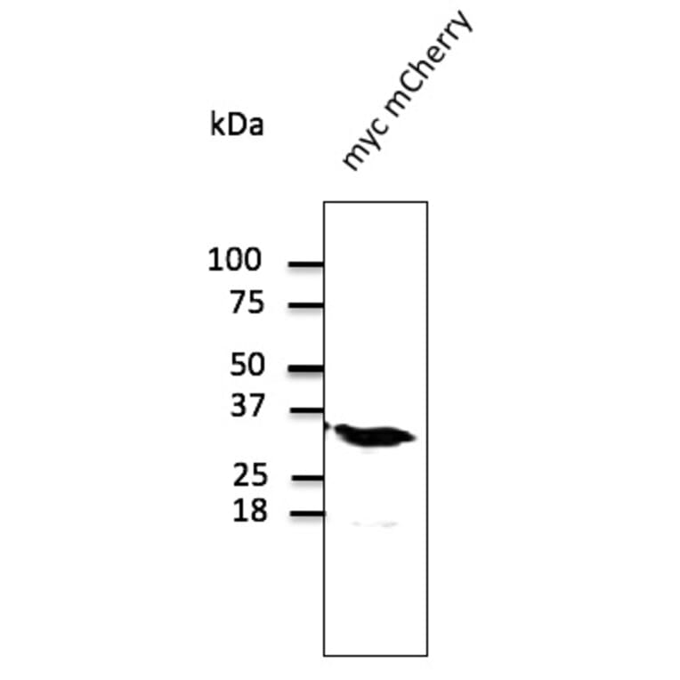
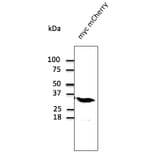
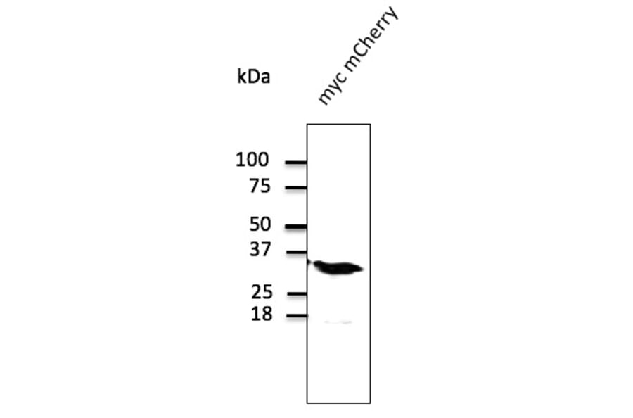
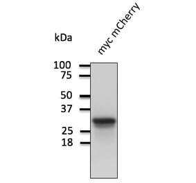
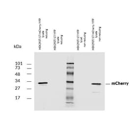
Western Blot - Anti-mCherry Antibody (A121571)
293 cells, transfected with myc-mCherry, detected with Anti-mCherry Antibody at a 1:1,000 dilution.
Enlarge Image
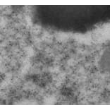
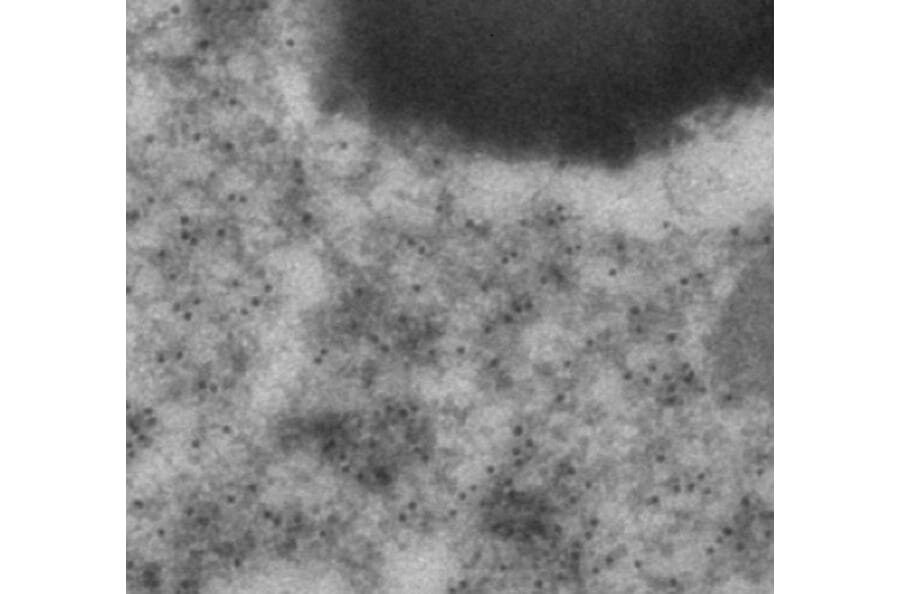
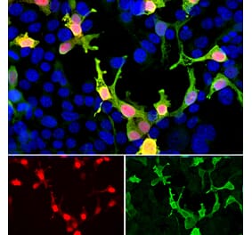
Anti-mCherry Antibody (A121571)
Immunogold labeling of RPE, in vivo injected with mCherry expressing vector, using Anti-mCherry Antibody.
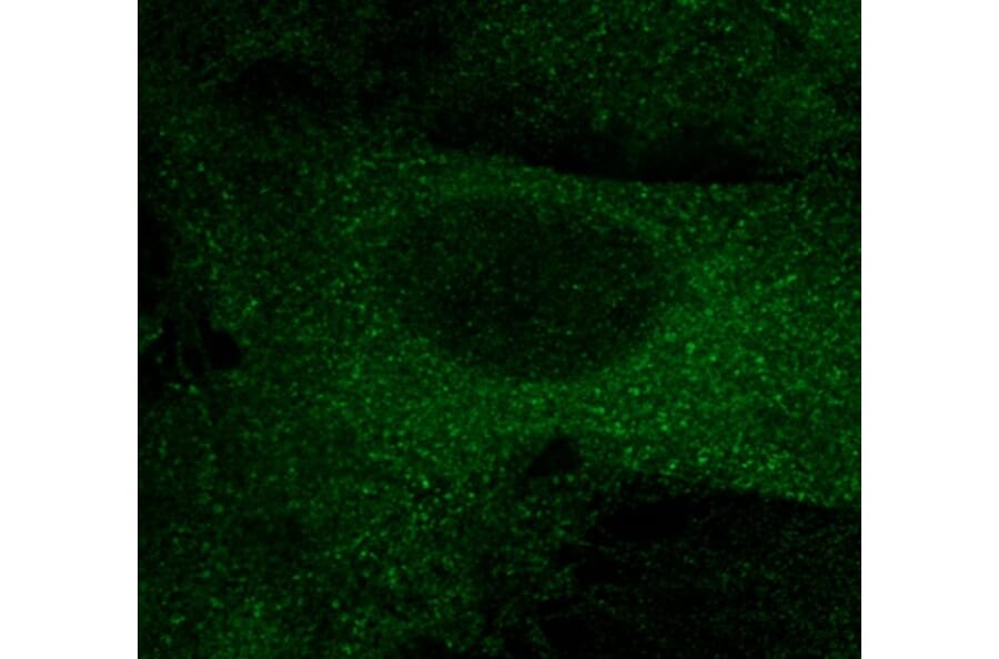
Enlarge Image
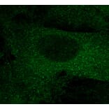
Anti-mCherry Antibody (A121571)
COS-7 cells, transfected with mCherry-EEA1, fixed with 4% PFA, stained with Anti-mCherry Antibody at a 1:100 dilution.
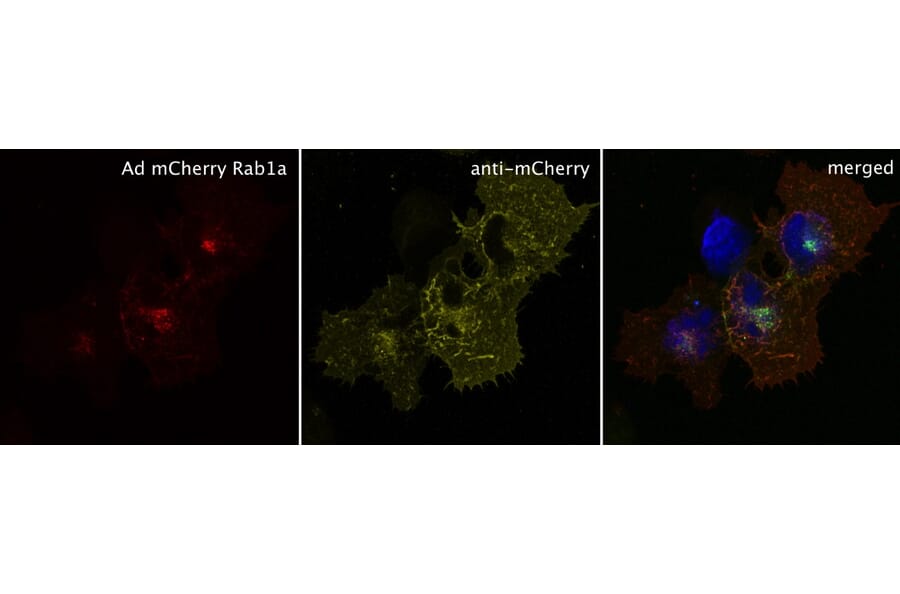
Enlarge Image
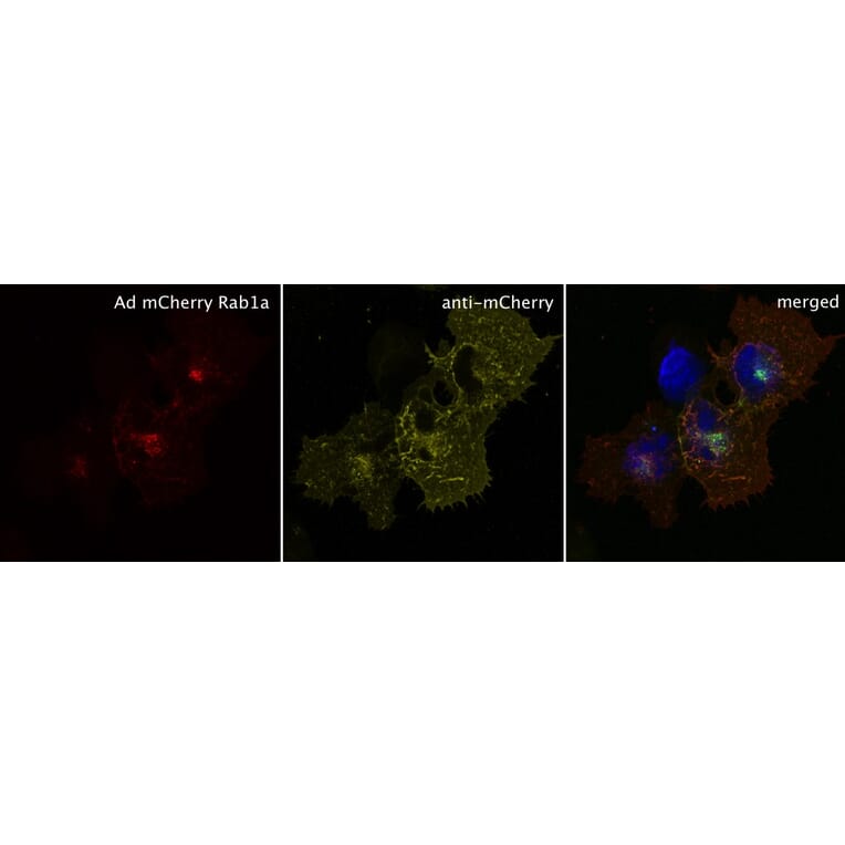
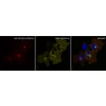
Anti-mCherry Antibody (A121571)
HEK293 cells, transduced with mCherry RAB1A, stained with Anti-mCherry Antibody.
Publishing research using Anti-mCherry Antibody (A121571)? Please
let us know
so that we can list the citation on this page.
Most popular Anti-mCherry Antibodies
(7)
A85306
4 Citations
Anti-mCherry Antibody
Rabbit polyclonal antibody to mCherry for WB, ICC/IF and IHC.
(5)
A85305
Anti-mCherry Antibody [1C51]
Mouse monoclonal [1C51] antibody to mCherry for WB, ICC/IF and IHC.
(5)
A85304
Anti-mCherry Antibody
Chicken polyclonal antibody to mCherry for WB, ICC/IF and IHC.
Alternative products to Anti-mCherry Antibody (A121571)
(5)
A121603
1 Citation
Anti-mCherry Antibody
Goat polyclonal antibody to mCherry for WB, IF, IHC-P, IHC-Fr and IEM.
(4)
A270547
Anti-mCherry Antibody
Goat polyclonal antibody to mCherry for WB, ICC/IF and IHC.
(4)
A104343
Anti-mCherry Antibody [5A6]
Mouse monoclonal [5A6] antibody to mCherry for WB, ICC/IF and IHC .
(3)
A285874
Anti-mCherry Antibody
Rabbit polyclonal antibody to mCherry for WB, ICC and Flow Cytometry.
A337467
Anti-mCherry Nanobody [SAA0406]
Recombinant mouse monoclonal [SAA0406] nanobody to mCherry for ELISA, ICC/IF and WB.
(2)
A337479
Anti-mCherry Nanobody [SAA1128]
Recombinant alpaca monoclonal [SAA1128] nanobody to mCherry for ELISA.
A1061
Anti-mCherry Antibody
Mouse monoclonal antibody to mCherry for WB.
A50025
Anti-mCherry Antibody
Mouse monoclonal antibody to mCherry for WB.
A337478
Anti-mCherry Nanobody [SAA0879]
Recombinant alpaca monoclonal [SAA0879] nanobody to mCherry for ELISA.
A322379
Anti-mCherry Antibody
Rabbit polyclonal antibody to mCherry for WB.
A322378
Anti-mCherry Antibody
Goat polyclonal antibody to mCherry for WB.
See all mCherry Antibodies
Top


![Immunohistochemistry - Anti-mCherry Antibody [1C51] (A85305) - Antibodies.com](https://cdn.antibodies.com/image/catalog/85/A85305_1.jpg?profile=product_alternative)

![Immunofluorescence - Anti-mCherry Antibody [5A6] (A104343) - Antibodies.com](https://cdn.antibodies.com/image/catalog/104/A104343_1.jpg?profile=product_alternative)
![Dose-response - Anti-mCherry Nanobody [SAA0406] (A337467) - Antibodies.com](https://cdn.antibodies.com/image/catalog/337/A337467_1.jpg?profile=product_alternative)
![SDS-PAGE - Anti-mCherry Nanobody [SAA1128] (A337479) - Antibodies.com](https://cdn.antibodies.com/image/catalog/337/A337479_1.jpg?profile=product_alternative)
![SDS-PAGE - Anti-mCherry Nanobody [SAA0879] (A337478) - Antibodies.com](https://cdn.antibodies.com/image/catalog/337/A337478_1.jpg?profile=product_alternative)
