Primary Antibodies
Secondary Antibodies
Proteins & Peptides
ELISA Kits
About Us
Contact Us
Sign In/Register
0
ISO 9001:2015 Certified
Live Customer Support
4.5/5 on Trustpilot
100% Quality Guarantee
Home
Primary Antibodies
MCP 1 Antibodies
Anti-MCP1 Antibody (A32636)
Anti-MCP1 Antibody (A32636)
Overview
Specifications
$720
Product Datasheet
Mouse monoclonal (S101) antibody to Monocyte Chemotactic Protein 1 (Biotin) for ELISA.
100% Guarantee
Price Match Guarantee
Product Size:
100µl
Quantity:
1
2
3
4
5
6
7
8
9
10
Add To Cart
Request a Quotation
Custom or Bulk Request
Shipping Information
Freight/Packing Charges:
$40
Dispatched from St. Louis, MO.
Lead Time: 6-9 business days.
Specifications
Name
Anti-MCP1 Antibody
Description
Mouse monoclonal (S101) antibody to Monocyte Chemotactic Protein 1 (Biotin).
Specificity
This antibody recognises natural and recombinant human MCP-1.
Applications
ELISA
Reactivity
Human
Cross Reactivity
This antibody does not recognise human IL-8, IL-1ß, SAA and EGF.
Immunogen
Purified recombinant human MCP-1.
Epitope
Sp2/0-Ag14
Host
Mouse
Clonality
Monoclonal
Clone ID
S101
Isotype
IgG1
Conjugate
Biotin
Purity
Protein G affinity chromatagraphy.
Product Form
Liquid
Formulation
Supplied in 0.01M Phosphate Buffered Saline, pH 7.2, with 1% Gelatin and 0.1% Proclin-300.
Storage
Shipped at 4°C. Upon delivery aliquot and store at -20°C. Avoid freeze / thaw cycles.
Synonyms
C-C motif chemokine 2, CCL2, HC11, MCAF, MCP-1, Monocyte chemoattractant protein 1, Monocyte chemotactic and activating factor, Monocyte chemotactic protein 1, Monocyte secretory protein JE, SCYA2, Small-inducible cytokine A2
Isotype Controls
Mouse IgG1 [MOPC-21] (Biotin) (A86204)
Disclaimer
This product is for research use only. It is not intended for diagnostic or therapeutic use.
Publishing research using Anti-MCP1 Antibody (A32636)? Please
let us know
so that we can list the citation on this page.
Alternative products to Anti-MCP1 Antibody (A32636)
A279615
Anti-MCP1 Antibody [AA03-4H10] (Biotin)
Mouse monoclonal [AA03-4H10] antibody to MCP1 (Biotin) for ELISA.
A282342
Anti-MCP1 Antibody (Biotin)
Goat polyclonal antibody to MCP1 (Biotin) for ELISA.
A325332
Anti-MCP1 Antibody [MTBH9] (Biotin)
Mouse monoclonal [MTBH9] antibody to MCP1 (Biotin) for ELISA.
A332071
Carlumab Biosimilar - Anti-MCP1 Antibody (Biotin)
Recombinant human monoclonal antibody to MCP1 (Biotin) for ELISA and Flow Cytometry.
Proteins predicted to interact with MCP 1
Predicted protein interactions based upon String database. Revelancy score correlates with probability of interaction.
RANTES Antibodies
99.9% Relevancy Score
MCP-2 Antibodies
99.9% Relevancy Score
Macrophage Inflammatory Protein 5 Antibodies
99.9% Relevancy Score
CCR5 Antibodies
99.9% Relevancy Score
IL-8 Antibodies
99.9% Relevancy Score
CXCL9 Antibodies
99.9% Relevancy Score
CCR1 Antibodies
99.9% Relevancy Score
CCR2 Antibodies
99.9% Relevancy Score
CXCL13 Antibodies
99.8% Relevancy Score
Top


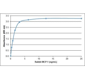
![ELISA - Anti-MCP1 Antibody [AA03-4H10] (Biotin) (A279615) - Antibodies.com](https://cdn.antibodies.com/image/catalog/279/A279615_1.jpg?profile=product_alternative)