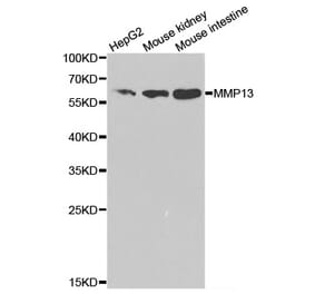
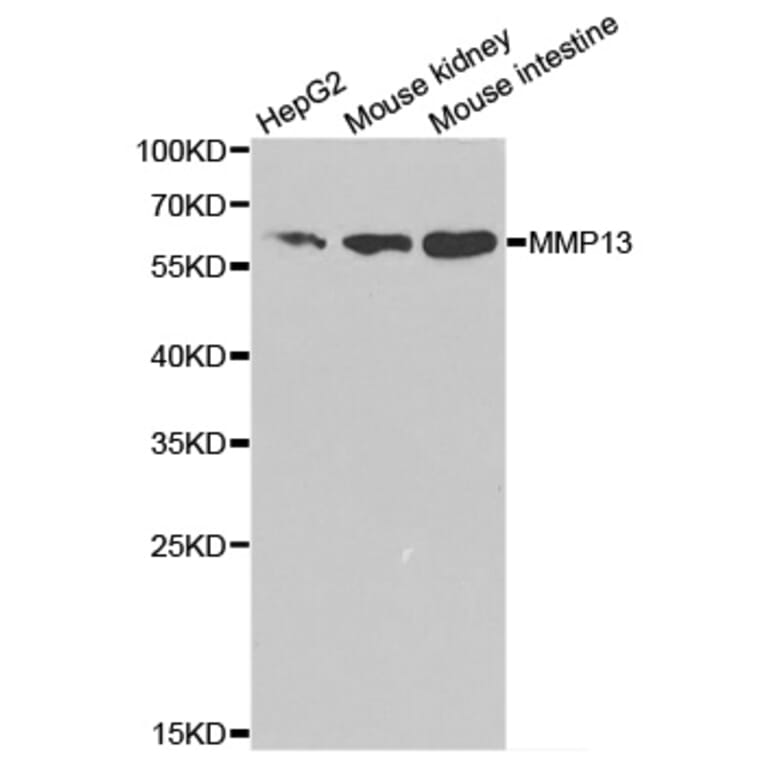
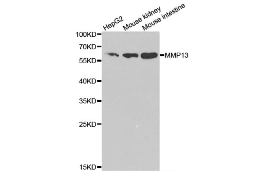
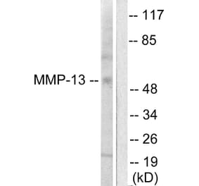
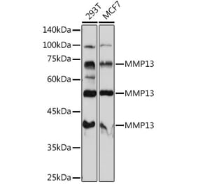
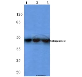
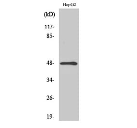
Unconjugated
To investigate the molecular pathogenesis of the canonical Wnt/β-catenin pathway in exercise-induced osteoarthritis (OA), 30 male healthy Sprague Dawley rats were divided into three groups (control, normal exercise‑induced OA and injured exercise‑induced OA groups) in order to establish the exercise‑induced OA rat model. The mRNA and protein expression levels of Runx‑2, BMP‑2, Ctnnb1, Sox‑9, collagen Ⅱ, Mmp‑13, Wnt‑3a and β‑catenin in chondrocytes were detected by reverse transcription‑quantitative polymerase chain reaction, western blotting and immunohistochemical staining. The mRNA levels of Runx‑2, BMP‑2 and Ctnnb1 were upregulated in the normal exercise‑induced OA and injured exercise‑induced OA groups; while Runx‑2 and BMP‑2 were upregulated in the injured exercise‑induced OA group when compared with the normal exercise‑induced OA group. The protein levels of Mmp‑13, Wnt‑3a and β‑catenin were increased and collagen Ⅱ was reduced in the normal exercise‑induced OA and injured exercise‑induced OA groups. Ctnnb1, Wnt‑3a and β‑catenin, which are key genes and proteins in the canonical Wnt/β‑catenin pathway, were abnormally expressed in chondrocytes of the exercise‑induced OA rat model. Ctnnb1, β‑catenin and Wnt‑3a were suggested to participate in the pathogenesis of exercise‑induced OA by abnormally activating the Wnt/β‑catenin pathway during physical exercise due to excessive pressure. The results of the present study may provide an improved understanding of the pathogenesis of exercise-induced OA.
OBJECTIVE:
In this study, we investigated the effects of hinokitiol on matrix metalloproteinase (MMP)-1, -3, -13, collagen type II (Col2a1) and β-catenin expressions in rat chondrocytes induced by interleukin-1β and in an experimental rat model induced by intra-articular injection of mono-iodoacetate (MIA) into the knee.
METHODS:
Chondrocytes were cultured from the articular cartilage of 2-week-old rats. Passaged chondrocytes were pretreated with hinokitiol for 2h followed by co-incubation with IL-1β for 24h. Quantitative real-time polymerase chain reaction and Western blotting were used to assess the expression of MMP-1, -3, -13, Col2a1 and β-catenin. Chondrocytes were also treated with Licl, Dickkopf-1, and/or hinokitiol for 24h, the MMP-1, -3, -13 and β-catenin protein levels determined by Western blotting. The in vivo effects of hinokitiol were assessed by morphological and histological analyses following MIA injection.
RESULTS:
Hinokitiol inhibited IL-1β-stimulated MMP-1,-3 and -13 expressions and IL-1β-induced activation of intracellular β-catenin proteins in cultured chondrocytes. In vivo, morphological and histological examinations demonstrated that hinokitiol significantly ameliorated cartilage degeneration.
CONCLUSIONS:
Hinokitiol is an effective anti-inflammatory reagent that acts by inhibiting the Wnt/β-catenin signaling pathway and could be a promising therapeutic agent for the prevention and treatment of osteoarthritis.
Copyright © 2014 Elsevier B.V. All rights reserved.