

- Primary Antibodies
- Secondary Antibodies
Fluorescent Conjugates
- Cyanine 3
- Cyanine 5
- Cyanine 5.5
- FITC
- PE
- Texas Red
- TRITC
- Unconjugated
Enzyme Conjugates
- Alkaline Phosphatase
- Biotin
- HRP
Application
- ELISA
- ICC/IF
- IHC
- Western Blot
- Proteins & Peptides
- ELISA Kits
- Custom Services
- Research Areas
- Customer SupportSupport


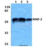
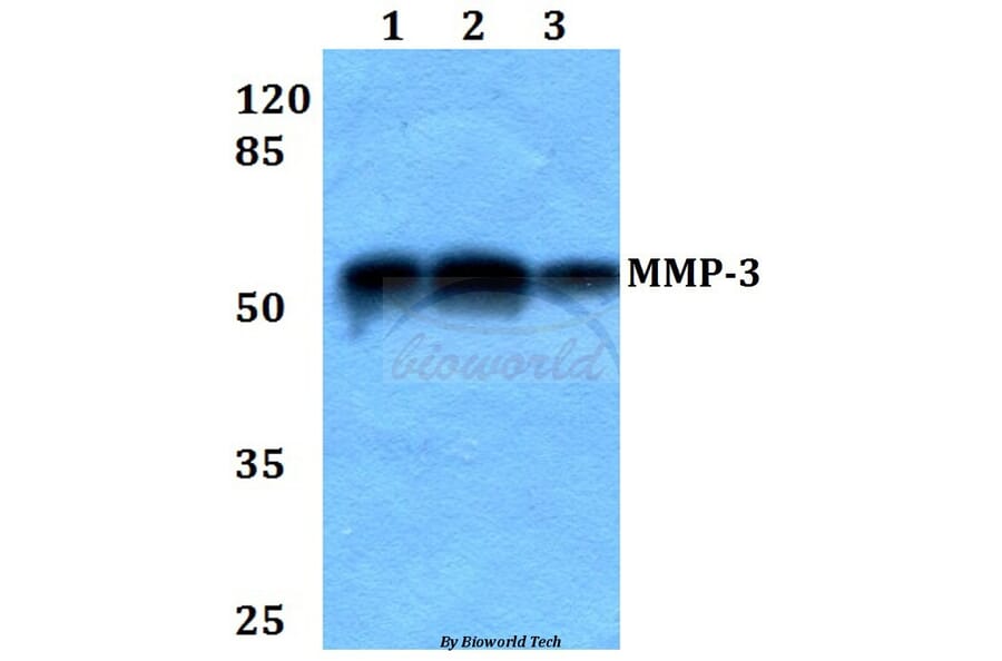


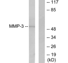
![Western Blot - Anti-MMP3 Antibody [ARC51098] (A309113) - Antibodies.com](https://cdn.antibodies.com/image/catalog/309/A309113_1.jpg?profile=product_alternative)
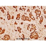
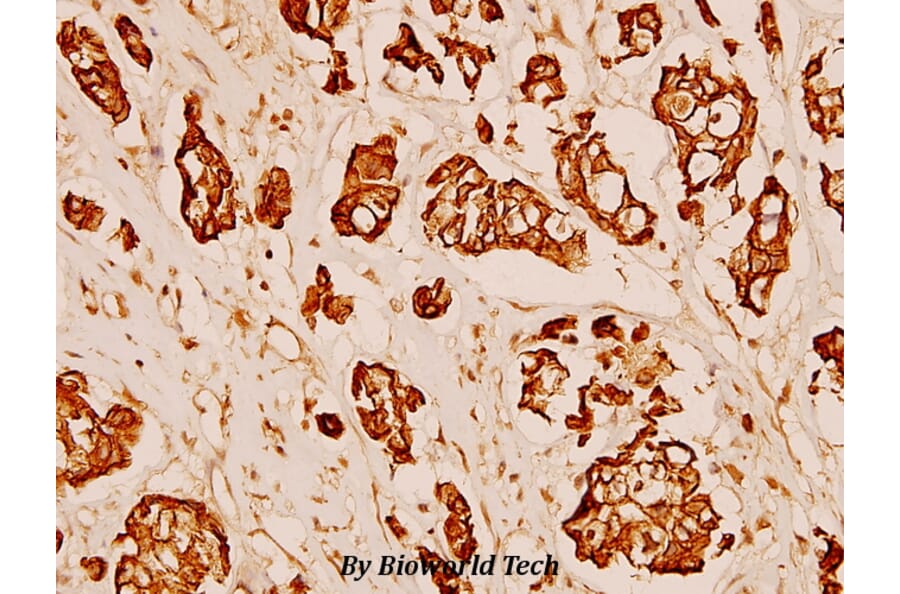
![Immunohistochemistry - Anti-MMP3 Antibody [MMP3/2806] (A249371) - Antibodies.com](https://cdn.antibodies.com/image/catalog/249/A249371_1.jpg?profile=product_alternative)
![Immunohistochemistry - Anti-MMP3 Antibody [MMP3/2806] - BSA and Azide free (A252551) - Antibodies.com](https://cdn.antibodies.com/image/catalog/252/A252551_1.jpg?profile=product_alternative)
![Western Blot - Anti-MMP3 Antibody [MMP3/2655] - BSA and Azide free (A252550) - Antibodies.com](https://cdn.antibodies.com/image/catalog/252/A252550_1.jpg?profile=product_alternative)
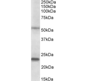
![SDS-PAGE - Anti-MMP3 Antibody [MMP3/1994R] (A249371) - Antibodies.com](https://cdn.antibodies.com/image/catalog/249/A249372_1.jpg?profile=product_alternative)
![SDS-PAGE - Anti-MMP3 Antibody [MMP3/1994R] - BSA and Azide free (A252551) - Antibodies.com](https://cdn.antibodies.com/image/catalog/252/A252552_1.jpg?profile=product_alternative)
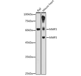
![Western Blot - Anti-MMP3 Antibody [MMP3/2655] (A249370) - Antibodies.com](https://cdn.antibodies.com/image/catalog/249/A249370_1.jpg?profile=product_alternative)
![Immunohistochemistry - Anti-MMP3 Antibody [rMMP3/1730] (A249368) - Antibodies.com](https://cdn.antibodies.com/image/catalog/249/A249369_1.jpg?profile=product_alternative)