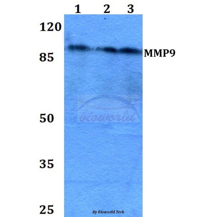
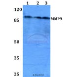
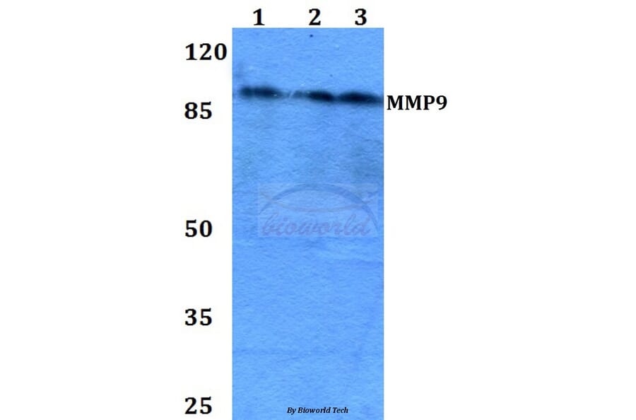
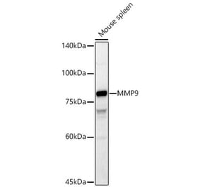
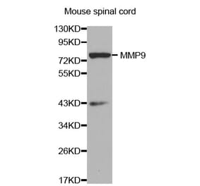
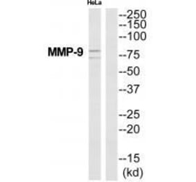
MMP9 (W680) pAb detects endogenous levels of MMP9 protein.
Applications
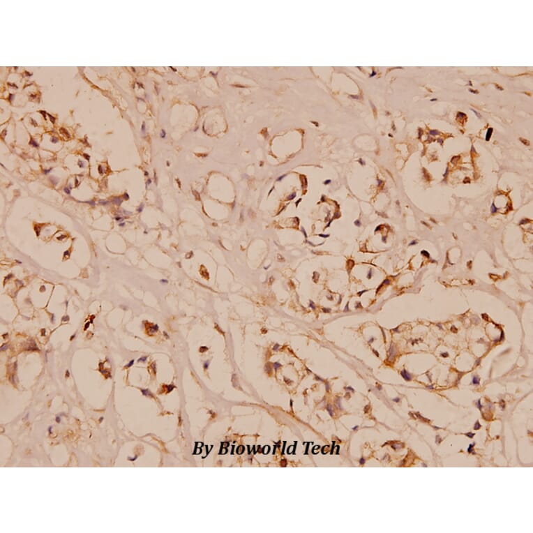
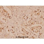
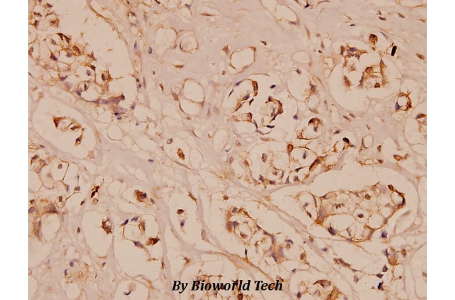
WB, IHC, IF
Reactivity
Human, Mouse, Rat
Immunogen
Synthetic peptide, corresponding to amino acids 650-700 of Human MMP9.
Host
Rabbit
Clonality
Polyclonal
Conjugate
Unconjugated
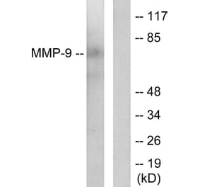
Molecular Weight
~ 92 kDa
Purity
The antibody was affinity-purified from rabbit antiserum by affinity-chromatography using epitope-specific immunogen and the purity is > 95% (by SDS-PAGE).
Product Form
1 mg/ml in Phosphate buffered saline (PBS) with 0.05% sodium azide, approx. pH 7.2.
Synonyms
92 kDa gelatinase, 92 kDa type IV collagenase, CLG4B, Gelatinase B, GELB, Matrix metalloproteinase-9, MMP-9
Recombinant chimeric monoclonal antibody to MMP9 for use as a research grade Andecaliximab biosimila for ELISA, Flow Cytometry, Functional Studies and in vivo Research.
Recombinant chimeric monoclonal antibody to MMP9 for use as a research grade Andecaliximab biosimila for ELISA, Flow Cytometry, Functional Studies and in vivo Research.


![Immunocytochemistry/Immunofluorescence - Anti-MMP9 Antibody [S51-82] (A305026) - Antibodies.com](https://cdn.antibodies.com/image/catalog/305/A305026_1.png?profile=product_alternative)
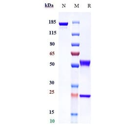
![SDS-PAGE - Anti-MMP9 Antibody [Research Grade Biosimilar] - Low endotoxin, Azide free (A324151) - Antibodies.com](https://cdn.antibodies.com/image/catalog/324/A324151_1.jpg?profile=product_alternative)

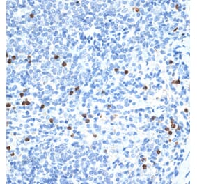
![Immunohistochemistry - Anti-MMP9 Antibody [2C3] (A252549) - Antibodies.com](https://cdn.antibodies.com/image/catalog/249/A249373_1.jpg?profile=product_alternative)