Figure 1: Western Blot - Anti-MSH2 Antibody [ARC55799] (A306655)
Western blot analysis of extracts from wild type(WT) and MSH2 knockout (KO) HeLa cells, using Anti-MSH2 Antibody [ARC55799] (A306655) at 1:20,000 dilution. The secondary antibody was Goat Anti-Rabbit IgG H&L Antibody (HRP) at 1:10,000 dilution. Lysates/proteins were present at 25µg per lane. The blocking buffer used was 3% non-fat dry milk in TBST. Detection was with a ECL Basic Kit. Exposure time: 60s.
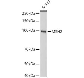
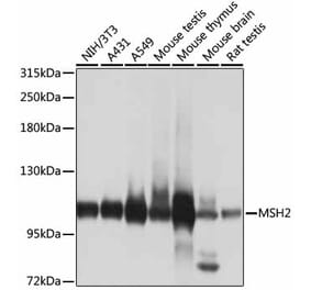
Figure 2: Western Blot - Anti-MSH2 Antibody [ARC55799] (A306655)
Western blot analysis of various lysates, using Anti-MSH2 Antibody [ARC55799] (A306655) at 1:20,000 dilution. The secondary antibody was Goat Anti-Rabbit IgG H&L Antibody (HRP) at 1:10,000 dilution. Lysates/proteins were present at 25µg per lane. The blocking buffer used was 3% non-fat dry milk in TBST. Detection was with a ECL Basic Kit. Exposure time: 60s.
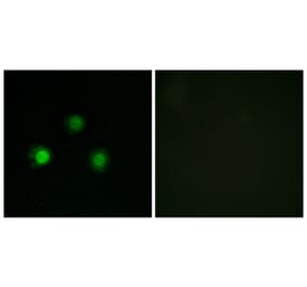
Immunohistochemistry analysis of paraffin-embedded human appendix tissue using Anti-MSH2 Antibody [ARC55799] (A306655) at a dilution of 1:100(40x lens). Perform high pressure antigen retrieval with 10 mM citrate buffer pH 6.0 before commencing with IHC staining protocol.
Immunohistochemistry analysis of paraffin-embedded human colon carcinoma tissue using Anti-MSH2 Antibody [ARC55799] (A306655) at a dilution of 1:100(40x lens). Perform high pressure antigen retrieval with 10 mM citrate buffer pH 6.0 before commencing with IHC staining protocol.
Immunohistochemistry analysis of paraffin-embedded human colon cancer loss of MSH2 expression using Anti-MSH2 Antibody [ARC55799] (A306655) at a dilution of 1:100(40x lens). Perform high pressure antigen retrieval with 10 mM citrate buffer pH 6.0 before commencing with IHC staining protocol.


![Western Blot - Anti-MSH2 Antibody [ARC55799] (A306655) - Antibodies.com](https://cdn.antibodies.com/image/catalog/306/A306655_1.jpg?profile=product_top)
![Western Blot - Anti-MSH2 Antibody [ARC55799] (A306655) - Antibodies.com](https://cdn.antibodies.com/image/catalog/306/A306655_2.jpg?profile=product_top)
![Immunohistochemistry - Anti-MSH2 Antibody [ARC55799] (A306655) - Antibodies.com](https://cdn.antibodies.com/image/catalog/306/A306655_3.jpg?profile=product_top)
![Immunohistochemistry - Anti-MSH2 Antibody [ARC55799] (A306655) - Antibodies.com](https://cdn.antibodies.com/image/catalog/306/A306655_4.jpg?profile=product_top)
![Immunohistochemistry - Anti-MSH2 Antibody [ARC55799] (A306655) - Antibodies.com](https://cdn.antibodies.com/image/catalog/306/A306655_5.jpg?profile=product_top)
![Western Blot - Anti-MSH2 Antibody [ARC55799] (A306655) - Antibodies.com](https://cdn.antibodies.com/image/catalog/306/A306655_1.jpg?profile=product_top_thumb)
![Western Blot - Anti-MSH2 Antibody [ARC55799] (A306655) - Antibodies.com](https://cdn.antibodies.com/image/catalog/306/A306655_2.jpg?profile=product_top_thumb)
![Immunohistochemistry - Anti-MSH2 Antibody [ARC55799] (A306655) - Antibodies.com](https://cdn.antibodies.com/image/catalog/306/A306655_3.jpg?profile=product_top_thumb)
![Immunohistochemistry - Anti-MSH2 Antibody [ARC55799] (A306655) - Antibodies.com](https://cdn.antibodies.com/image/catalog/306/A306655_4.jpg?profile=product_top_thumb)
![Immunohistochemistry - Anti-MSH2 Antibody [ARC55799] (A306655) - Antibodies.com](https://cdn.antibodies.com/image/catalog/306/A306655_5.jpg?profile=product_top_thumb)
![Western Blot - Anti-MSH2 Antibody [ARC55799] (A306655) - Antibodies.com](https://cdn.antibodies.com/image/catalog/306/A306655_1.jpg?profile=product_image)
![Western Blot - Anti-MSH2 Antibody [ARC55799] (A306655) - Antibodies.com](https://cdn.antibodies.com/image/catalog/306/A306655_2.jpg?profile=product_image)
![Immunohistochemistry - Anti-MSH2 Antibody [ARC55799] (A306655) - Antibodies.com](https://cdn.antibodies.com/image/catalog/306/A306655_3.jpg?profile=product_image)
![Immunohistochemistry - Anti-MSH2 Antibody [ARC55799] (A306655) - Antibodies.com](https://cdn.antibodies.com/image/catalog/306/A306655_4.jpg?profile=product_image)
![Immunohistochemistry - Anti-MSH2 Antibody [ARC55799] (A306655) - Antibodies.com](https://cdn.antibodies.com/image/catalog/306/A306655_5.jpg?profile=product_image)
![Immunohistochemistry - Anti-MSH2 Antibody [MSH2/2622] (A249381) - Antibodies.com](https://cdn.antibodies.com/image/catalog/249/A249381_1.jpg?profile=product_alternative)
![Immunohistochemistry - Anti-MSH2 Antibody [MSH2/2622] - BSA and Azide free (A252561) - Antibodies.com](https://cdn.antibodies.com/image/catalog/252/A252561_1.jpg?profile=product_alternative)
![Western Blot - Anti-MSH2 Antibody [ARC1285] (A307085) - Antibodies.com](https://cdn.antibodies.com/image/catalog/307/A307085_1.jpg?profile=product_alternative)
![Immunohistochemistry - Anti-MSH2 Antibody [rMSH2/6548] (A249380) - Antibodies.com](https://cdn.antibodies.com/image/catalog/249/A249380_1.jpg?profile=product_alternative)
![Immunohistochemistry - Anti-MSH2 Antibody [rMSH2/6548] - BSA and Azide free (A252560) - Antibodies.com](https://cdn.antibodies.com/image/catalog/252/A252560_1.jpg?profile=product_alternative)
![Immunohistochemistry - Anti-MSH2 Antibody [MSH2/6549R] (A249382) - Antibodies.com](https://cdn.antibodies.com/image/catalog/249/A249382_1.jpg?profile=product_alternative)
![Immunohistochemistry - Anti-MSH2 Antibody [MSH2/6549R] - BSA and Azide free (A252562) - Antibodies.com](https://cdn.antibodies.com/image/catalog/252/A252562_1.jpg?profile=product_alternative)
![Immunohistochemistry - Anti-MSH2 Antibody [MSH2/6852] (A277717) - Antibodies.com](https://cdn.antibodies.com/image/catalog/277/A277717_1.jpg?profile=product_alternative)
![Immunohistochemistry - Anti-MSH2 Antibody [MSH2/6852] - BSA and Azide free (A278305) - Antibodies.com](https://cdn.antibodies.com/image/catalog/278/A278305_1.jpg?profile=product_alternative)