Primary Antibodies
Secondary Antibodies
Proteins & Peptides
ELISA Kits
About Us
Contact Us
Sign In/Register
0
ISO 9001:2015 Certified
Live Customer Support
4.5/5 on Trustpilot
100% Quality Guarantee
Home
Primary Antibodies
RAB27A Antibodies
Anti-RAB27A Antibody (A121563)
Anti-RAB27A Antibody (A121563)
Overview
Specifications
Images
Enlarge Image
Enlarge Image
Enlarge Image
$590
Product Datasheet
Goat polyclonal antibody to RAB27A for WB, IF, IHC-P and IHC-Fr.
100% Guarantee
Price Match Guarantee
Product Size:
200µg
500µg
Quantity:
1
2
3
4
5
6
7
8
9
10
Add To Cart
Request a Quotation
Custom or Bulk Request
Shipping Information
Freight/Packing Charges:
$40
Dispatched from St. Louis, MO.
Lead Time: 5-8 business days.
Specifications
Name
Anti-RAB27A Antibody
Description
Goat polyclonal antibody to RAB27A.
Specificity
This antibody recognizes RAB27A and RAB27B.
Applications
WB
,
IF
,
IHC-P
,
IHC-Fr
Dilutions
WB: 1:250-1:2,000, IF: 1:50-1:200, IHC-P: 1:50-1:400, IHC-Fr: 1:50-1:400
Reactivity
Human, Rat, Mouse, Canine, Monkey
Immunogen
Recombinant peptide derived from within residues a.a. 120 to the C terminus of mouse RAB27A, expressed in and purified from E. coli.
Host
Goat
Clonality
Polyclonal
Isotype
IgG
Conjugate
Unconjugated
Purification
Affinity purification.
Concentration
1 mg/ml
Product Form
Liquid
Formulation
Supplied in Phosphate Buffered Saline with 20% Glycerol and 0.05% Sodium Azide.
Storage
Shipped at 4°C. Upon delivery aliquot and store at -20°C. Avoid freeze/thaw cycles.
Synonyms
GTP-binding protein Ram, Rab-27, RAB27, Ras-related protein Rab-27A
Isotype Controls
Goat IgG (A121671)
Suitable Secondaries
Donkey Anti-Goat IgG H&L Antibody (AP) (A300679)
Donkey Anti-Goat IgG H&L Antibody (Biotin) (A300716)
Donkey Anti-Goat IgG H&L Antibody (FITC) (A300685)
Donkey Anti-Goat IgG H&L Antibody (HRP) (A300730)
See all Anti-Goat IgG Secondaries →
Disclaimer
This product is for research use only. It is not intended for diagnostic or therapeutic use.
Scientific Validation Data
Validation Data
(3)
Enlarge Image
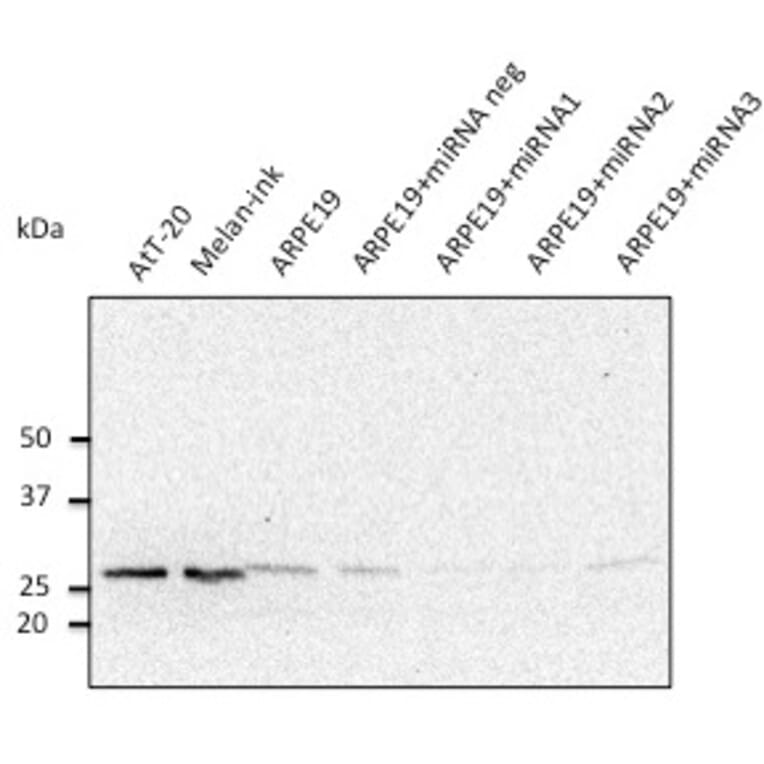
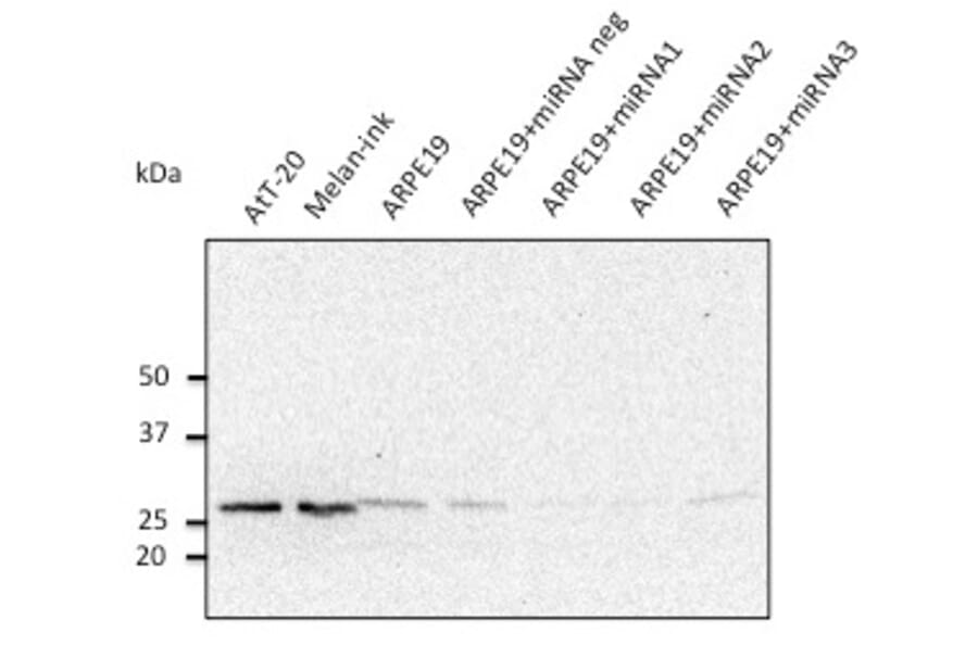
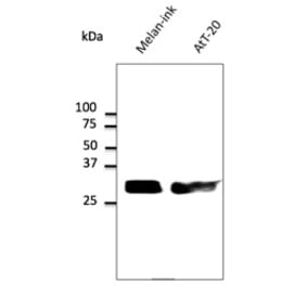
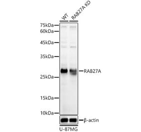
Western Blot - Anti-RAB27A Antibody (A121563)
Various lysates detected with Anti-RAB27A Antibody at a 1:500 dilution. Rabbit anti-goat IgG antibody (HRP) was used at a 1:10,000 dilution.
Enlarge Image
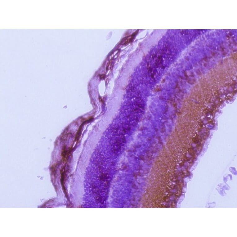
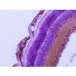
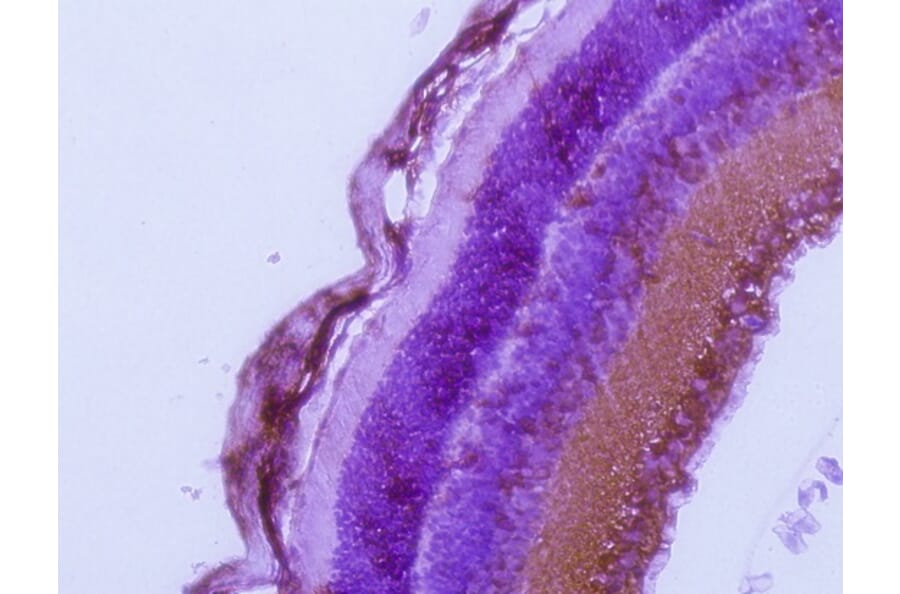
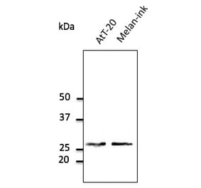
Immunohistochemistry - Anti-RAB27A Antibody (A121563)
Anti-RAB27A Antibody (1:200) staining of formalin-fixed, paraffin-embedded albino mouse retina, after heat-induced antigen retrieval.
Enlarge Image
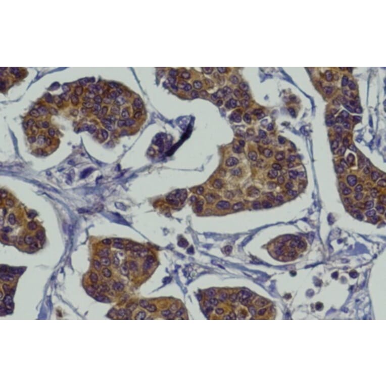
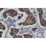
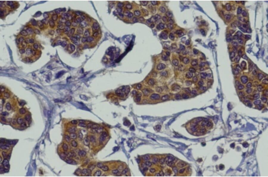
Immunohistochemistry - Anti-RAB27A Antibody (A121563)
Anti-RAB27A Antibody (1:200) staining of formalin-fixed, paraffin-embedded mammary tissue, after heat-induced antigen retrieval.
Publishing research using Anti-RAB27A Antibody (A121563)? Please
let us know
so that we can list the citation on this page.
Alternative products to Anti-RAB27A Antibody (A121563)
(5)
A121688
Anti-RAB27 Antibody
Goat polyclonal antibody to RAB27 for WB, IF, IHC-P and IHC-Fr.
(4)
A121575
Anti-RAB27A Antibody
Goat polyclonal antibody to RAB27A for WB, IF, IHC-P and IHC-Fr.
(3)
A37040
Anti-RAB27A Antibody
Rabbit polyclonal antibody to RAB27A for WB and ICC/IF.
A30525
Anti-RAB27A Antibody
Rabbit polyclonal antibody to RAB27A for WB and IHC.
(2)
A13754
Anti-RAB27A Antibody
Rabbit polyclonal antibody to RAB27A for WB and ICC/IF.
A348071
Anti-RAB27A Antibody [25GB1245] kappa
Mouse monoclonal [25GB1245] antibody to RAB27A for WB.
A42390
Anti-RAB27A Antibody
Rabbit polyclonal antibody to RAB27A for WB.
A348070
Anti-RAB27A Antibody [24GB7165] kappa
Mouse monoclonal [24GB7165] antibody to RAB27A for WB.
A348069
Anti-RAB27A Antibody [24GB8550]
Mouse monoclonal [24GB8550] antibody to RAB27A for WB.
See all RAB27A Antibodies
Proteins predicted to interact with RAB27A
Predicted protein interactions based upon String database. Revelancy score correlates with probability of interaction.
Granuphilin Antibodies
99.9% Relevancy Score
MYRIP Antibodies
99.9% Relevancy Score
MYO5A Antibodies
99.9% Relevancy Score
UNC13D Antibodies
99.9% Relevancy Score
Melanophilin Antibodies
99.9% Relevancy Score
Rabphilin 3A Antibodies
94.7% Relevancy Score
SYTL5 Antibodies
93.6% Relevancy Score
CHM Antibodies
93.5% Relevancy Score
STX11 Antibodies
93.1% Relevancy Score
Top