
| Sample Type | n | Range | Average |
|---|---|---|---|
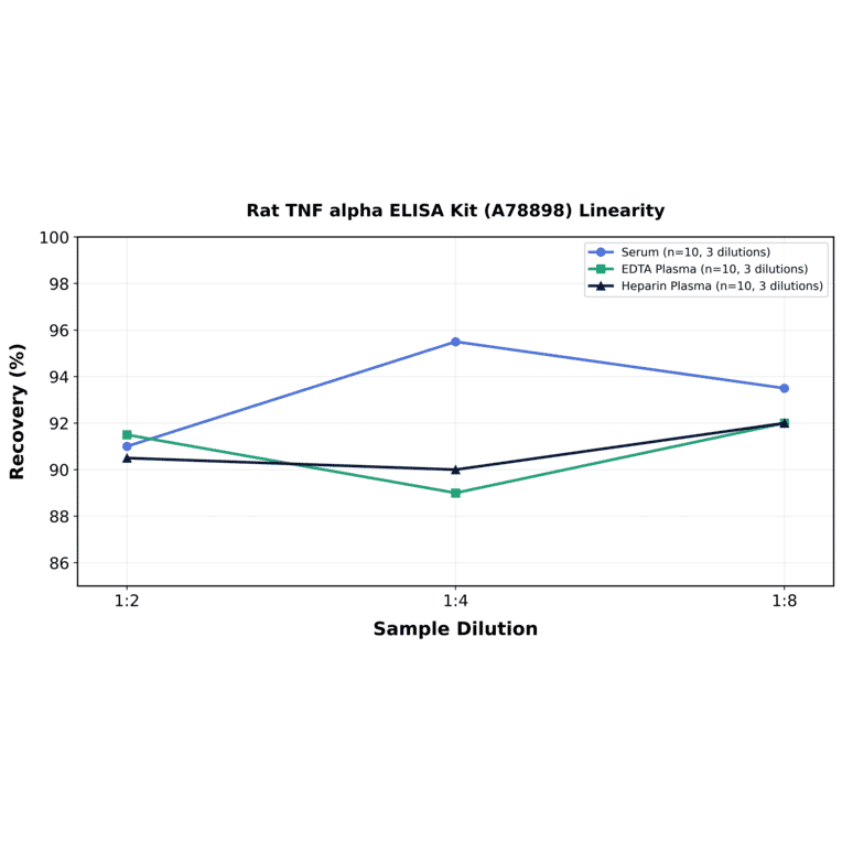
| Serum | 10 | 88% - 96% | 93% |
| EDTA Plasma | 10 | 90% - 97% | 94% |
| Heparin Plasma | 10 | 86% - 104% | 96% |
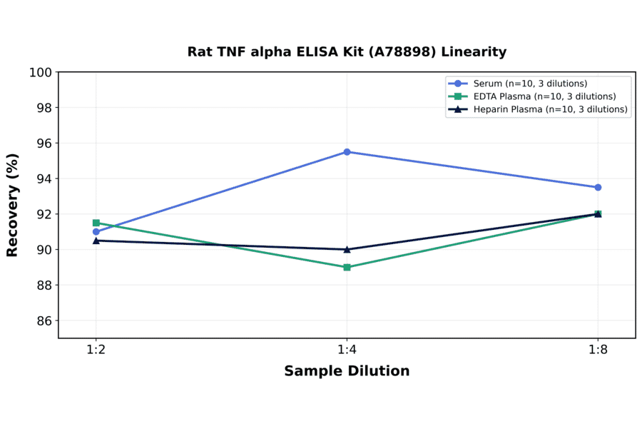
| Sample Type | n | 1:2 | 1:4 | 1:8 |
|---|---|---|---|---|
| Serum | 10 | 89-93% | 86-105% | 87-100% |
| EDTA Plasma | 10 | 85-98% | 82-96% | 83-101% |
| Heparin Plasma | 10 | 81-100% | 81-99% | 84-100% |
| Item | Quantity | Storage |
|---|---|---|
| Pre-Coated 96 Well Microplate | 12 x 8 Well Strips | +4°C |
| Lyopholized Standard | 2 Vials | +4°C |
| Sample Dilution Buffer | 20ml | +4°C |
| Biotinylated Detection Antibody | 120µl | +4°C |
| Antibody Dilution Buffer | 10ml | +4°C |
| HRP-Streptavidin Conjugate | 120µl | +4°C |
| SABC Dilution Buffer | 10ml | +4°C |
| TMB Substrate | 10ml | +4°C |
| Stop Solution | 10ml | +4°C |
| Wash Buffer (25X) | 30ml | +4°C |
| Plate Sealers | 5 Adhesive Strips | - |
| Foil Pouch | 1 Zip-Sealed Pouch | - |
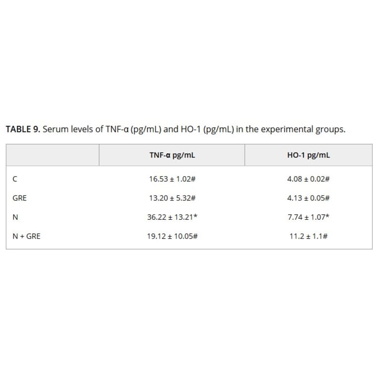
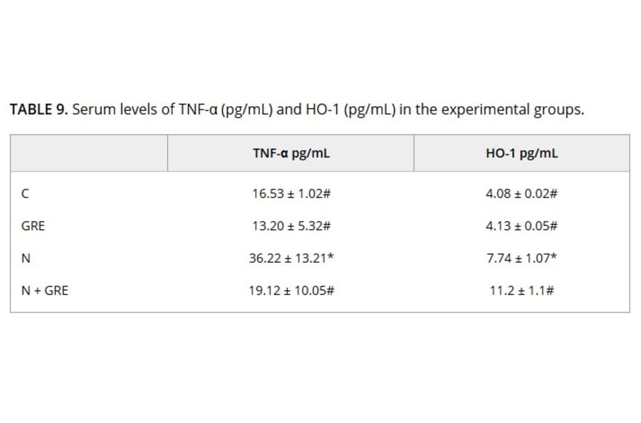
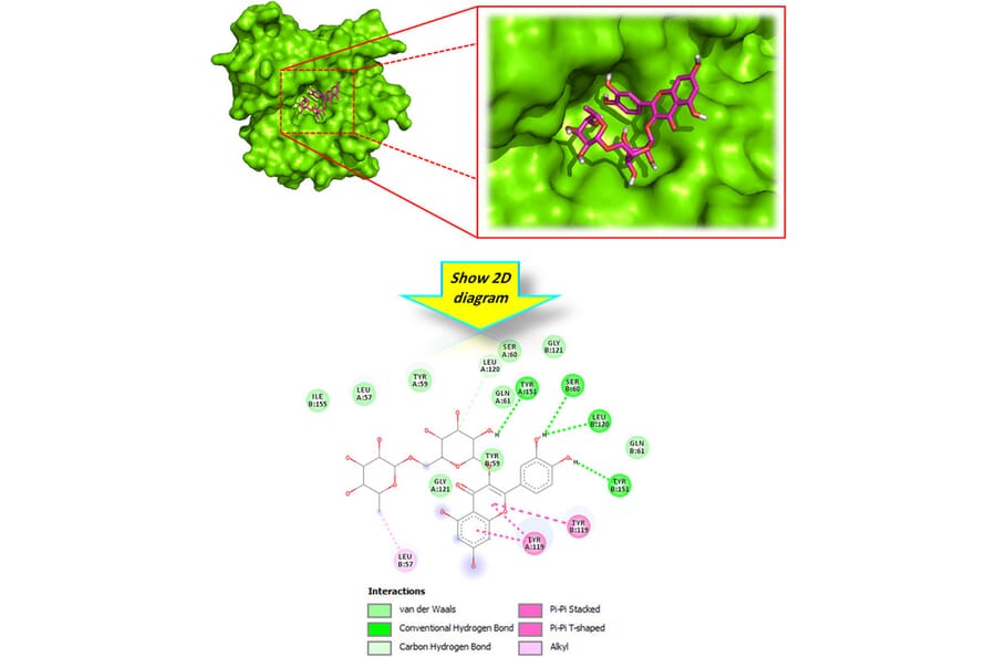
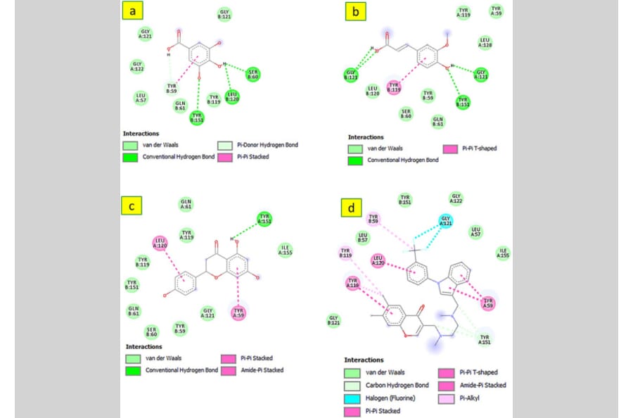
This study evaluated the cardioprotective effects of Alpinia galanga rhizome extract (GRE) against noise-induced myocardial injury via phytochemical profiling, molecular docking, and in vivo assessment. Male Wistar rats (n = 6/group) were assigned to the following four groups: control (C), control + GRE (100 mg/kg), noise-exposed (N), and noise-exposed + GRE (N+GRE, 100 mg/kg). Rats in the N and N+GRE groups were exposed to 90 dB(A) white noise for 2 h/day for 28 days, with GRE administered orally throughout the exposure period. Phytochemical analysis confirmed the presence of flavonoids and phenolic acids with known antioxidant and anti-inflammatory activities. In vitro, GRE significantly reduced nitric oxide production in lipopolysaccharide-stimulated RAW264.7 macrophages. In vivo, noise exposure elevated cardiac malondialdehyde levels, impaired antioxidant enzyme activity, and increased circulating tumor necrosis factor-alpha (TNF-a) and heme oxygenase-1 (HO-1) levels. GRE treatment restored redox balance, suppressed proinflammatory mediator levels, and improved histopathological alterations. Molecular docking analysis indicated strong binding of GRE phytoconstituents to HO-1 and TNF-a, supporting the observed in vivo effects. These findings demonstrate that GRE mitigates noise-induced cardiac injury through its antioxidant and anti-inflammatory properties, highlighting its therapeutic potential.
Background: In addition to their primary lipid-lowering effects, statins also exhibit pleiotropic properties, including anti-inflammatory, immunomodulatory, antimicrobial, antioxidative, and angiogenic effects, all of which promote wound healing. Rosuvastatin, a synthetic hydrophilic statin, has recently been proposed to enhance wound healing by stimulating angiogenesis and accelerating tissue regeneration. Its hydrophilic nature, longer half-life, greater hepatoselectivity, and better efficacy/safety than other statins are believed to contribute to its superior efficacy in wound repair, as suggested by promising preliminary results. However, current data on its use remain limited, necessitating further preclinical and clinical studies to thoroughly investigate this novel treatment option for burns.
Aim: To investigate the effects of rosuvastatin and its mechanism of action on burn wound healing process in an experimental study.
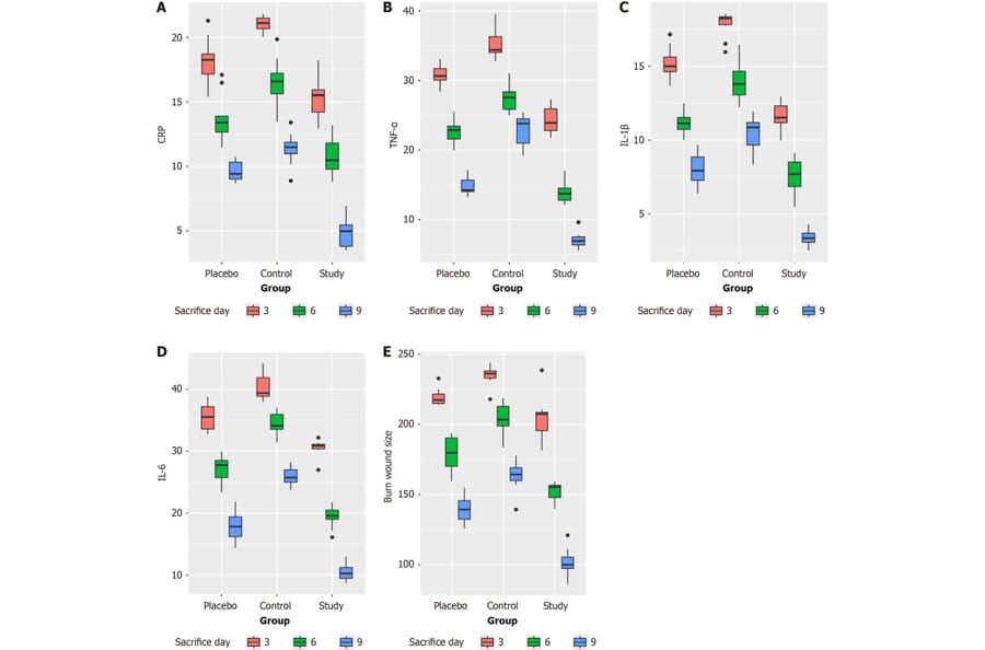
Methods: Ninety male Wistar albino rats aged 12-16 weeks were randomly assigned to three groups of 30, subjected to burn using specific stainless steel sealer. Burn eschar was removed the following day applying topical rosuvastatin cream (study), Eucerin cream (placebo), normal saline (control), and sterile wound dressing. Each group was divided into three subgroups of ten according to sacrifice day (3rd, 6th, 9th). C-reactive protein (CRP), tumor necrosis factor-alpha (TNF-a), interleukin (IL)-1ß, IL-6, digital assessment of burn healing, and histopathology were performed.
Results: Using the value on the third day in the control group as the baseline, reductions were measured on the sixth and ninth days. Similarly, reduction values were recorded on the third, sixth, and ninth days in the study group. A statistically significant reduction was observed in the study group compared to the control group, with greater reductions corresponding to later sacrifice days (3rd, 6th, and 9th) in CRP (P < 0.01), TNF-a (P < 0.01), IL-1ß (P < 0.01), and IL-6 (P < 0.01) levels and burn size (P < 0.01). Histopathology revealed a statistically significant reduction in inflammatory infiltration, coagulative necrosis, and microhemorrhage (P < 0.01). Conversely, statistically significant increases in neovascularization (P = 0.012) and fibroblastic reactions (P = 0.023) were noted. No adverse effects or deaths were observed.
Conclusion: Rosuvastatin reduces inflammation by lowering TNF-a, IL-1ß, IL-6, CRP while increasing neo-angiogenesis, fibroblast reactions and microenvironmental protection in burn wounds. These effects promote repair and positively impact burn wound healing.