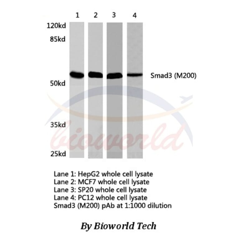
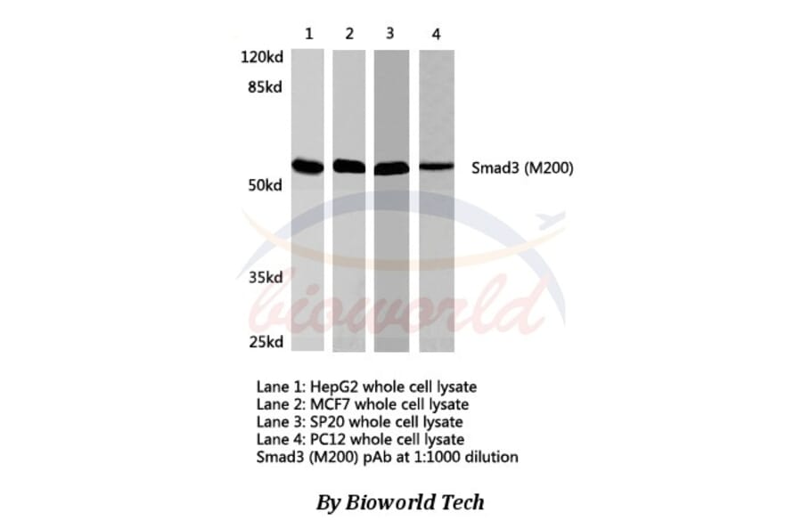
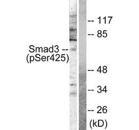
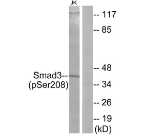
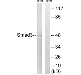
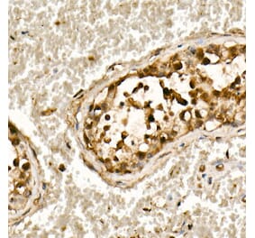
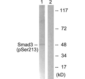
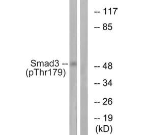
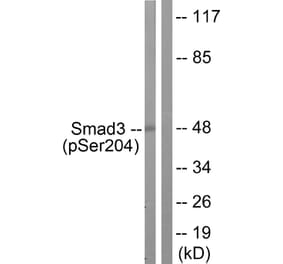
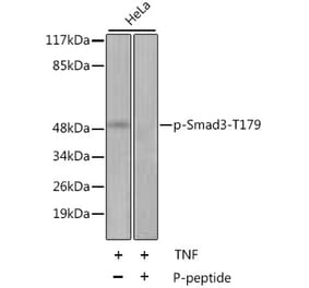
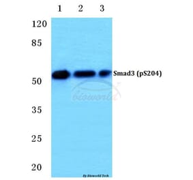
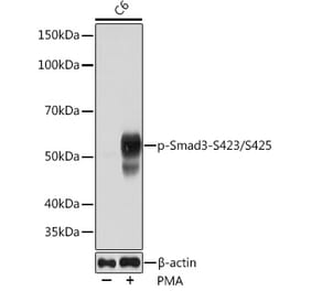
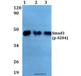
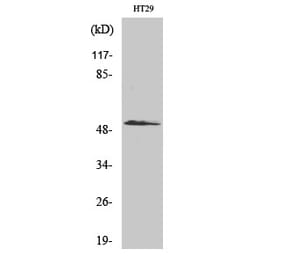
Unconjugated
Previous studies have shown that Astragalus and Paeoniae Radix Rubra extract (APE) is capable of protecting against liver fibrosis in rats. The hypothesis of the present study was that APE exerts its anti‑fibrotic effect by mediating the transforming growth factor β (TGF‑β)/Smad signaling pathway. In order to investigate this hypothesis, a series of assays were designed to detect the effects of APE on cell proliferation, cell invasion and the activation of hepatic stellate cells (HSCs). In addition, the effects of APE on the TGF‑β/Smad signaling pathway were explored, with the aim of elucidating the underlying mechanisms. HSCs were initially isolated from normal rat liver. A number of assays were then employed in order to evaluate the effects of APE on the function of these cells. Cell proliferation was investigated using an MTT assay and cell invasion was observed with the use of transwell invasion chambers. Collagen synthesis was measured with a 3H‑proline incorporation assay and expression of α‑smooth muscle actin was used to determine the extent of HSC activation. Protein expression induced by TGF‑β1 in HSCs was investigated by western blot and immunofluorescence analyses. Plasminogen activator inhibitor type1 (PAI‑1) and urokinase‑type plasminogen activator (uPA) transcriptional activity was measured using reverse transcription polymerase chain reaction. The results demonstrated that APE (5‑80 µg/ml) significantly inhibited fetal bovine serum‑induced cell proliferation in a dose‑dependent manner. Cell invasion and activation of HSCs induced by TGF‑β1 were disrupted by treatment with APE in a dose‑dependent manner. TGF‑β1 was observed to increase the phosphorylation of Smad2/3, while APE administered at higher doses produced inhibitory effects on Smad2/3 phosphorylation. In addition, administration of APE abrogated the TGF‑β1‑induced reduction in Smad‑7 expression in a dose‑dependent manner. The results further indicated that APE treatment not only reduced PAI‑1 expression, but also increased uPA expression in a dose‑dependent manner. In conclusion, APE exerted inhibitory effects on cell proliferation, invasion and activation of HSCs, and the mechanisms underlying these effects may involve the TGF‑β1/Smad pathway.
Human Dachshund homologue 1 (DACH1) is a major component of the Retinal Determination Gene Network. Loss of DACH1 expression was found in breast, prostate, lung, endometrial, colorectal and hepatocellular carcinoma. To explore the expression, regulation and function of DACH1 in human esophageal cancer, 11 esophageal cancer cell lines, 10 cases of normal esophageal mucosa, 51 cases of different grades of dysplasia and 104 cases of primary esophageal squamous cancer were employed. Methylation specific PCR, immunohistochemistry, western blot, flow cytometry, small interfering RNAs, colony formation techniques and xenograft mice model were used. We found that DACH1 expression was regulated by promoter region hypermethylation in esophageal cancer cell lines. 18.8% (6 of 32) of grade 1, 42.1% (8 of 19) of grade 2 and grade 3 dysplasia (ED2,3), and 61.5% (64 of 104) of esophageal cancer were methylated, but no methylation was found in 10 cases of normal esophageal mucosa. The methylation was increased in progression tendency during esophageal carcinogenesis (P<0.01). DACH1 methylation was associated with poor differentiation (P<0.05) and late tumor stage (P<0.05). Restoration of DACH1 expression inhibited cell growth and activated TGF-β signaling in KYSE150 and KYSE510 cells. DACH1 suppressed human esophageal cancer cell tumor growth in xenograft mice. In conclusion, DACH1 is frequently methylated in human esophageal cancer and methylation of DACH1 is involved in the early stage of esophageal carcinogenesis. DACH1 expression is regulated by promoter region hypermethylation. DACH1 suppresses esophageal cancer growth by activating TGF-β signaling.