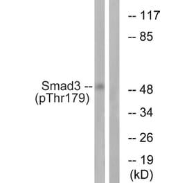
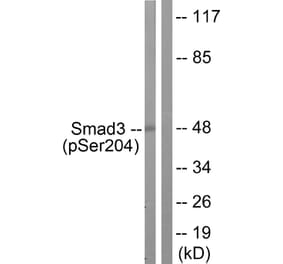
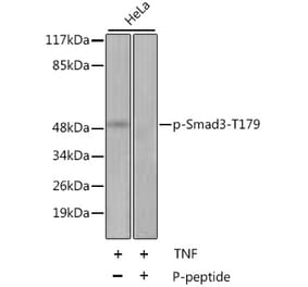
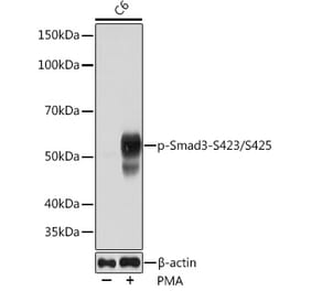
Rabbit polyclonal antibody to Smad3 (phospho-S204)
Specificity
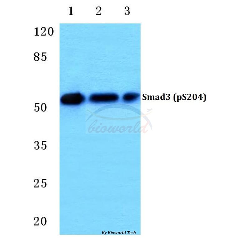
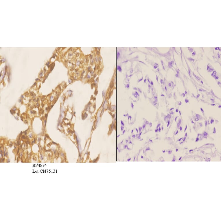
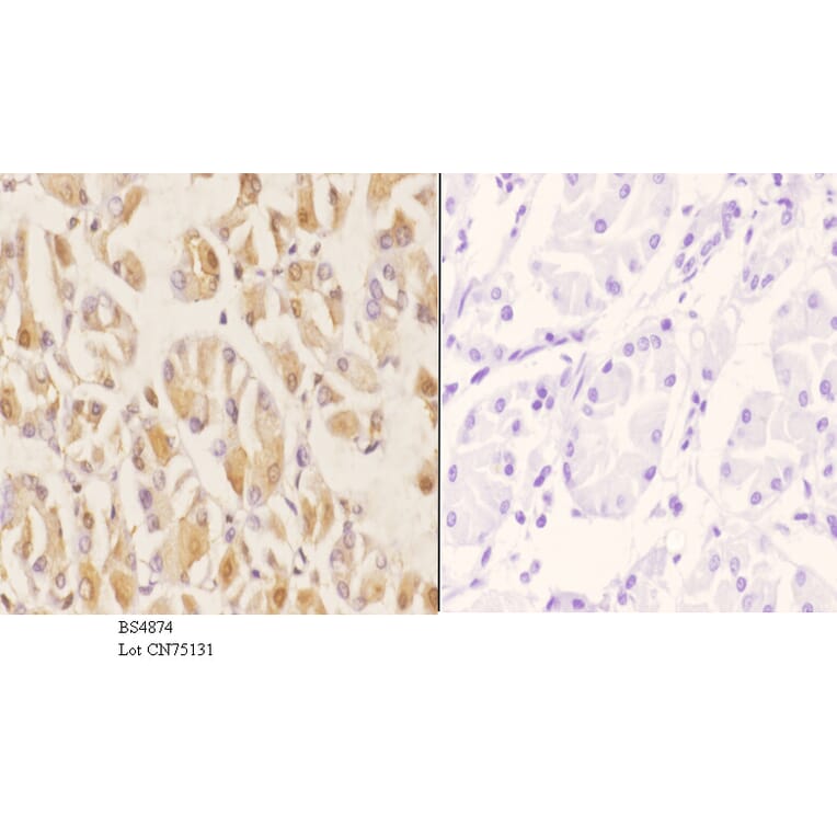
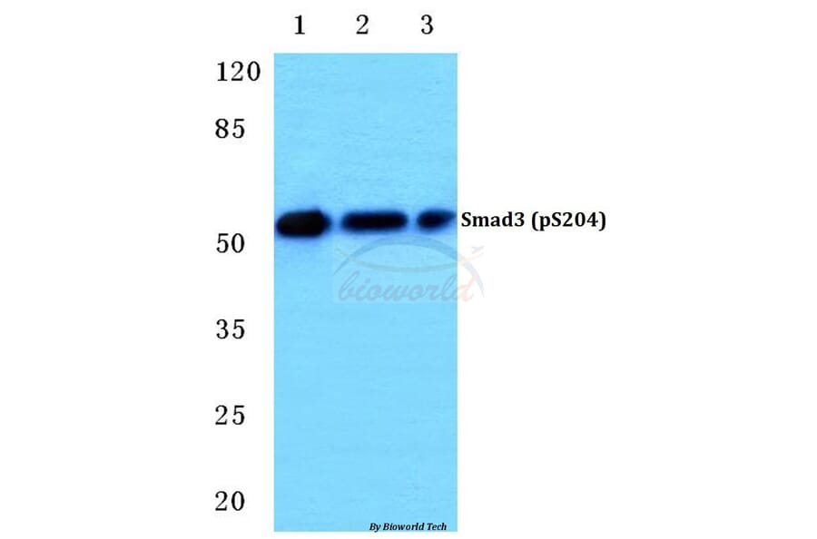
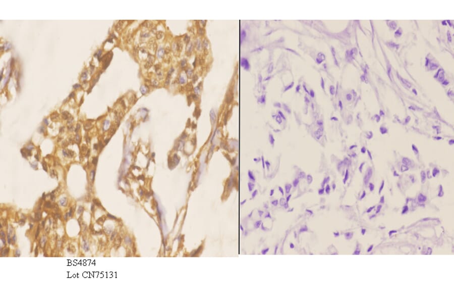
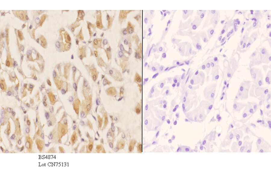
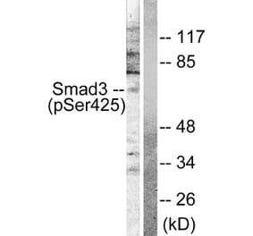
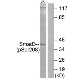
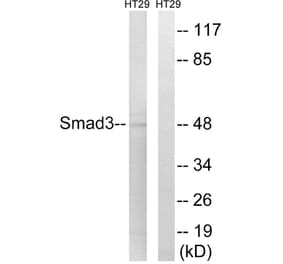
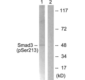
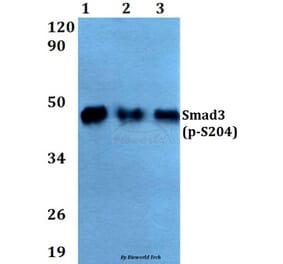
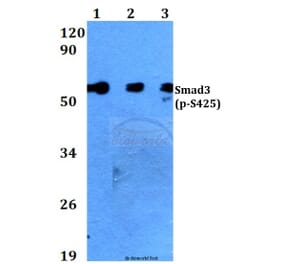
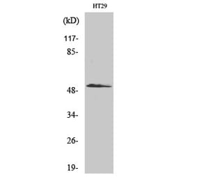
p-Smad3 (S204) pAb detects endogenous levels of Smad3 protein only when phosphorylated at Ser204.
Applications
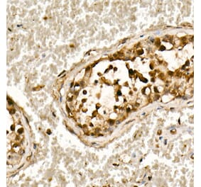
WB, IHC, ICC, IP
Reactivity
Human, Mouse, Rat
Immunogen
Synthetic phosphopeptide derived from human Smad3 around the phosphorylation site of Serine 204.
Host
Rabbit
Clonality
Polyclonal
Conjugate
Unconjugated
Molecular Weight
~ 48, 55 kDa
Purity
The antibody was affinity-purified from rabbit antiserum by affinity-chromatography using epitope-specific immunogen and the purity is > 95% (by SDS-PAGE).
Product Form
1 mg/ml in Phosphate buffered saline (PBS) with 15 mM sodium azide, approx. pH 7.2.
Synonyms
hMAD-3, hSMAD3, JV15-2, MAD homolog 3, Mad3, MADH3, Mothers against decapentaplegic homolog 3, Mothers against DPP homolog 3, SMAD 3, SMAD family member 3