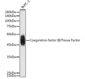
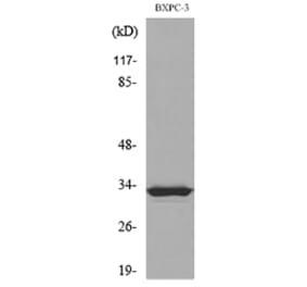
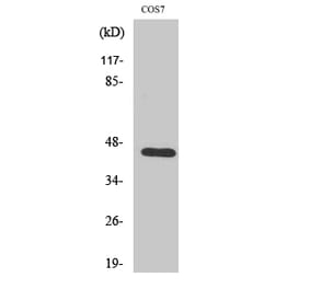
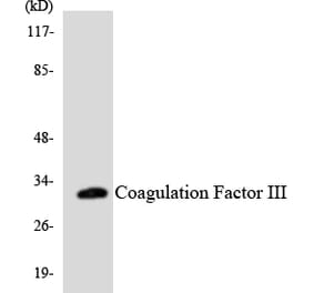
Figure 1: Western Blot - Anti-Tissue Factor Antibody [ARC60559] (A309523)
Western blot analysis of various lysates, using Anti-Tissue Factor Antibody [ARC60559] (A309523) at 1:1,000 dilution. The secondary antibody was Goat Anti-Rabbit IgG H&L Antibody (HRP) at 1:10,000 dilution. Lysates/proteins were present at 25µg per lane. The blocking buffer used was 3% non-fat dry milk in TBST. Detection was with a ECL Basic Kit. Exposure time: 10s.
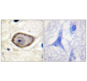
Immunohistochemistry analysis of paraffin-embedded human lung cancer using Anti-Tissue Factor Antibody [ARC60559] (A309523) at a dilution of 1:400 (40x lens). Perform high pressure antigen retrieval with 10 mM citrate buffer pH 6.0 before commencing with IHC staining protocol.
Immunofluorescence analysis of A-431 and MCF7(Negative sample) cells using Anti-Tissue Factor Antibody [ARC60559] (A309523) at a dilution of 1:300 (40x lens). DAPI was used to stain the cell nuclei (blue).
Publishing research using Anti-Tissue Factor Antibody [ARC60559] (A309523)? Please let us know so that we can list the citation on this page.
Alternative products to Anti-Tissue Factor Antibody [ARC60559] (A309523)


![Western Blot - Anti-Tissue Factor Antibody [ARC60559] (A309523) - Antibodies.com](https://cdn.antibodies.com/image/catalog/309/A309523_1.jpg?profile=product_top)
![Immunohistochemistry - Anti-Tissue Factor Antibody [ARC60559] (A309523) - Antibodies.com](https://cdn.antibodies.com/image/catalog/309/A309523_2.jpg?profile=product_top)
![Immunofluorescence - Anti-Tissue Factor Antibody [ARC60559] (A309523) - Antibodies.com](https://cdn.antibodies.com/image/catalog/309/A309523_3.jpg?profile=product_top)
![Western Blot - Anti-Tissue Factor Antibody [ARC60559] (A309523) - Antibodies.com](https://cdn.antibodies.com/image/catalog/309/A309523_1.jpg?profile=product_top_thumb)
![Immunohistochemistry - Anti-Tissue Factor Antibody [ARC60559] (A309523) - Antibodies.com](https://cdn.antibodies.com/image/catalog/309/A309523_2.jpg?profile=product_top_thumb)
![Immunofluorescence - Anti-Tissue Factor Antibody [ARC60559] (A309523) - Antibodies.com](https://cdn.antibodies.com/image/catalog/309/A309523_3.jpg?profile=product_top_thumb)
![Western Blot - Anti-Tissue Factor Antibody [ARC60559] (A309523) - Antibodies.com](https://cdn.antibodies.com/image/catalog/309/A309523_1.jpg?profile=product_image)
![Immunohistochemistry - Anti-Tissue Factor Antibody [ARC60559] (A309523) - Antibodies.com](https://cdn.antibodies.com/image/catalog/309/A309523_2.jpg?profile=product_image)
![Immunofluorescence - Anti-Tissue Factor Antibody [ARC60559] (A309523) - Antibodies.com](https://cdn.antibodies.com/image/catalog/309/A309523_3.jpg?profile=product_image)

![ELISA - Anti-Tissue Factor Antibody [Tisotumab Biosimilar] - Azide free (A318845) - Antibodies.com](https://cdn.antibodies.com/image/catalog/318/A318845_1.jpg?profile=product_alternative)
![Immunohistochemistry - Anti-Tissue Factor Antibody [IHC517] (A324501) - Antibodies.com](https://cdn.antibodies.com/image/catalog/324/A324501_1.jpg?profile=product_alternative)
![Flow Cytometry - Anti-Tissue Factor Chimeric Antibody [DMC463] - Azide free (A318727) - Antibodies.com](https://cdn.antibodies.com/image/catalog/318/A318727_1.jpg?profile=product_alternative)