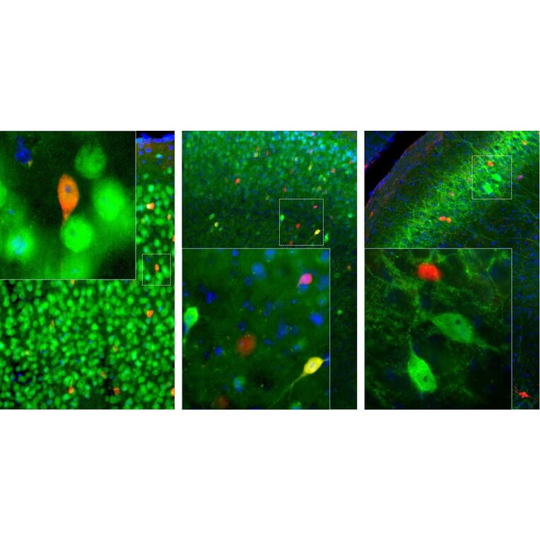
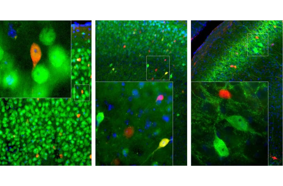
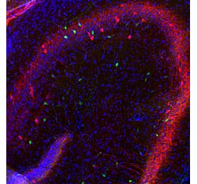
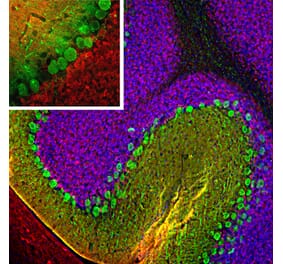
Left: Adult mouse brain section (45 µM; fixed by transcardial perfusion with 4% paraformaldehyde) across motor cortex was co-stained with Anti-Calretinin Antibody (red) and Anti-Fox3/NeuN Antibody (A85404 | green). Middle: Adult mouse brain section across visual cortex was co-stained with Anti-Calretinin Antibody (red) and Anti-Calbindin Antibody (A85359 | green). Calretinin and calbindin label different population of neurons in the brain. As a result, most cells were labeled with one of the two antibodies and appear to be either red or green. However in visual cortex, a few cells express both proteins and appear to be yellow. Right: Adult rat brain section (45 µM; fixed by transcardial perfusion with 4% paraformaldehyde) across hippocampal CA1 region was co-stained with Anti-Calretinin Antibody (red) and Anti-Parvalbumin Antibody (A85317 | green). The two antibodies stain distinct subsets of interneurons in the pyramidal layer and the positively labeled cells appear to be either red or green. Insets show high magnification pictures of boxed are in each image. Blue is a Hoechst staining that labels DNA.
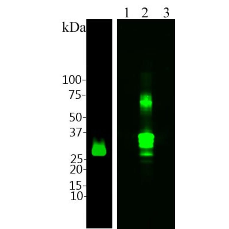
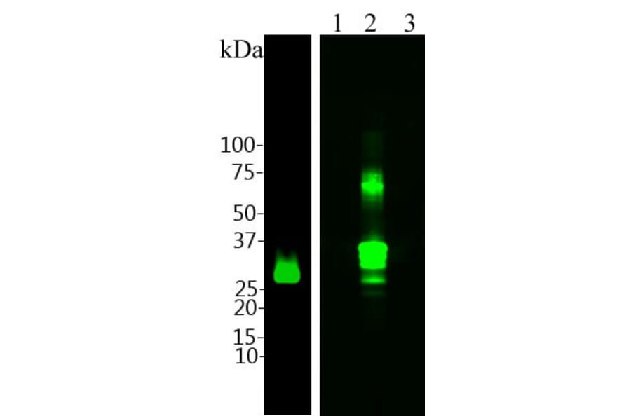
Left: Blot of 20µg of rat brain lysates probed with Anti-Calretinin Antibody (1:5,000). The antibody recognizes a clear band at ~29 kDa as expected. Right: At the same antibody concentration we probed a blot on which were 0.2µg of recombinant human parvalbumin (Lane 1), recombinant human calretinin (Lane 2) and recombinant human calbindin (Lane 3). Anti-Calretinin Antibody reacts only with the calretinin protein, and not the two related proteins parvalbumin and calbindin.
Alternative products to Anti-Calretinin Antibody (A85364)


![Immunofluorescence - Anti-Calretinin Antibody [6A9] (A85366) - Antibodies.com](https://cdn.antibodies.com/image/catalog/85/A85366_1.jpg?profile=product_alternative)
![Immunofluorescence - Anti-Calretinin Antibody [3G9] (A85367) - Antibodies.com](https://cdn.antibodies.com/image/catalog/85/A85367_1.jpg?profile=product_alternative)
![Immunohistochemistry - Anti-Calretinin Antibody [CALB2/2602] (A250372) - Antibodies.com](https://cdn.antibodies.com/image/catalog/250/A250372_1.jpg?profile=product_alternative)
![Immunohistochemistry - Anti-Calretinin Antibody [CALB2/2786] - BSA and Azide free (A253555) - Antibodies.com](https://cdn.antibodies.com/image/catalog/253/A253555_1.jpg?profile=product_alternative)
![Immunohistochemistry - Anti-Calretinin Antibody [CALB2/2807] (A250376) - Antibodies.com](https://cdn.antibodies.com/image/catalog/250/A250376_1.jpg?profile=product_alternative)
![Immunohistochemistry - Anti-Calretinin Antibody [CALB2/2786] (A250375) - Antibodies.com](https://cdn.antibodies.com/image/catalog/250/A250375_1.jpg?profile=product_alternative)
![Immunohistochemistry - Anti-Calretinin Antibody [CALB2/2602] - BSA and Azide free (A253552) - Antibodies.com](https://cdn.antibodies.com/image/catalog/253/A253552_1.jpg?profile=product_alternative)