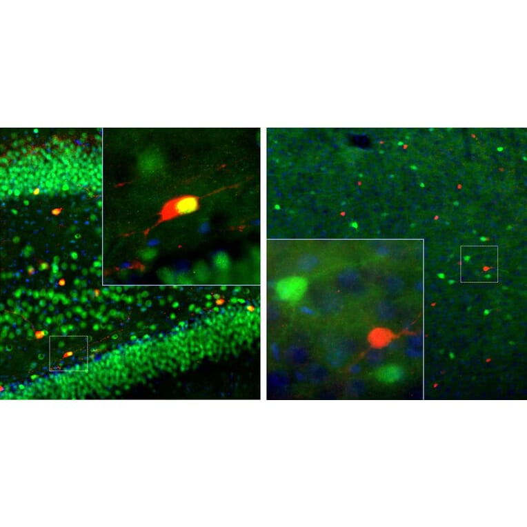
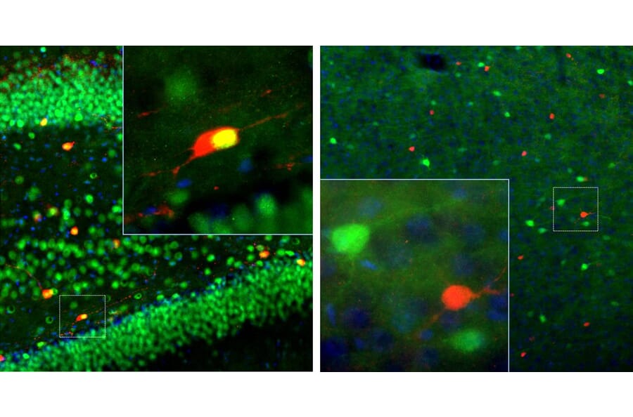
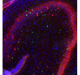
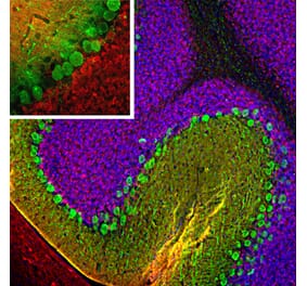
Left: Adult rat brain hippocampus section (45 µM; fixed by transcardial perfusion with 4% paraformaldehyde) was stained with Anti-Calretinin Antibody (1:1,000 | red) and Anti-MeCP2 Antibody (A85427 | 1,1000 | green). Anti-Calretinin Antibody labels a subset of hippocampal interneurons, which also express MeCP2 in the nucleus to give a yellow color. Right: Adult rat cortex section was co-stained with Anti-Calretinin Antibody (red) and Anti-Calbindin Antibody (A85360 | green). Each antibody specifically labels a subset of interneurons (i.e., calretinin-positive or calbindin-postive) that express each marker exclusively. Insets are high-magnification images of the boxed area in each picture. Blue is DAPI staining that labels DNA.
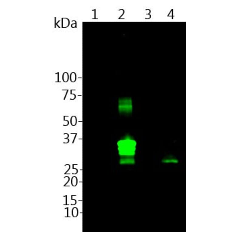
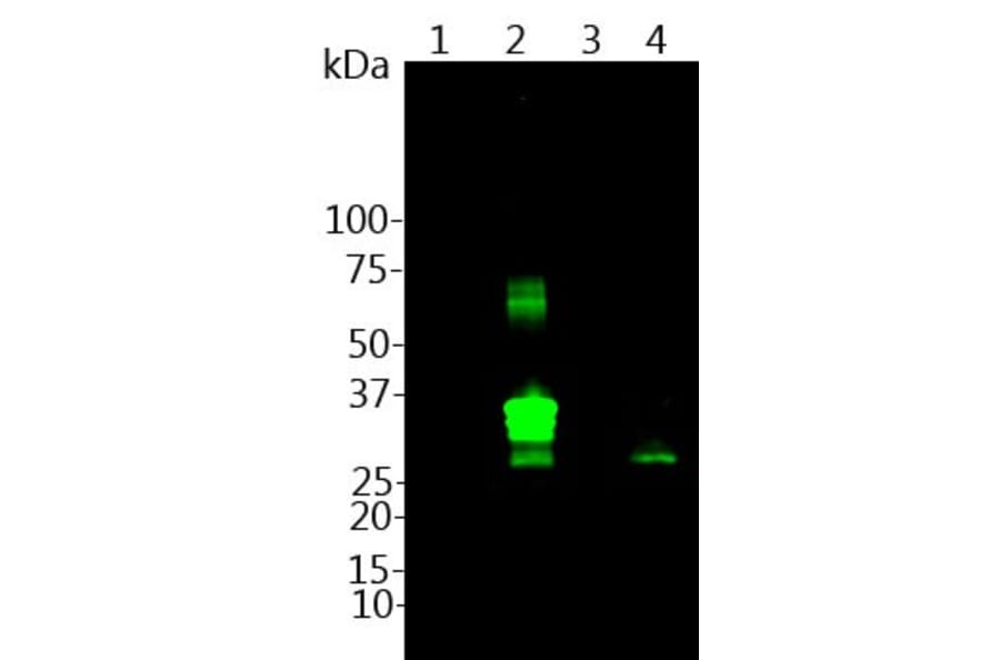
Western blot of recombinant protein parvalbumin (Lane 1), calretinin (Lane 2), calbindin (Lane 3) and rat brain lysates (Lane 4 ) was probed with Anti-Calretinin Antibody (1:1,000). In rat brain lysates, this antibody recognizes a clear band at ~29 kDa and it reacts only with calretinin protein, and not other calcium-binding proteins.
Alternative products to Anti-Calretinin Antibody (A85365)


![Immunofluorescence - Anti-Calretinin Antibody [6A9] (A85366) - Antibodies.com](https://cdn.antibodies.com/image/catalog/85/A85366_1.jpg?profile=product_alternative)
![Immunofluorescence - Anti-Calretinin Antibody [3G9] (A85367) - Antibodies.com](https://cdn.antibodies.com/image/catalog/85/A85367_1.jpg?profile=product_alternative)
![Immunohistochemistry - Anti-Calretinin Antibody [CALB2/2602] (A250372) - Antibodies.com](https://cdn.antibodies.com/image/catalog/250/A250372_1.jpg?profile=product_alternative)
![Immunohistochemistry - Anti-Calretinin Antibody [CALB2/2602] - BSA and Azide free (A253552) - Antibodies.com](https://cdn.antibodies.com/image/catalog/253/A253552_1.jpg?profile=product_alternative)
![Immunohistochemistry - Anti-Calretinin Antibody [CALB2/2807] - BSA and Azide free (A253556) - Antibodies.com](https://cdn.antibodies.com/image/catalog/253/A253556_1.jpg?profile=product_alternative)
![Immunohistochemistry - Anti-Calretinin Antibody [CALB2/2786] - BSA and Azide free (A253555) - Antibodies.com](https://cdn.antibodies.com/image/catalog/253/A253555_1.jpg?profile=product_alternative)
![Immunohistochemistry - Anti-Calretinin Antibody [CALB2/2807] (A250376) - Antibodies.com](https://cdn.antibodies.com/image/catalog/250/A250376_1.jpg?profile=product_alternative)