CF® Dye and biotin conjugates: Add 0.5 ml dH2O for reconstitution. HRP or DNP conjugates: Add 1 ml dH2O for reconstitution.
Formulation
Supplied in Phosphate Buffered Saline containing 50% glycerol, 2 mg/ml BSA, and 0.05% Sodium Azide.
Storage
Shipped at +4°C. Upon delivery aliquot and store at -20°C. Avoid freeze/thaw cycles. This product is also photosensitive and should be protected from light. Should this product contain a precipitate we recommend microcentrifugation before use. CF® Dyes are guaranteed for at least 6 months from data of receipt when stored correctly.
General Notes
Looking for a specific protein conjugate to simplify your workflow? We offer a library of over 2,000 targets conjugated to your choice of CF® dye. To enquire about a custom product, contact us directly.
Disclaimer
This product is for research use only. It is not intended for diagnostic or therapeutic use.
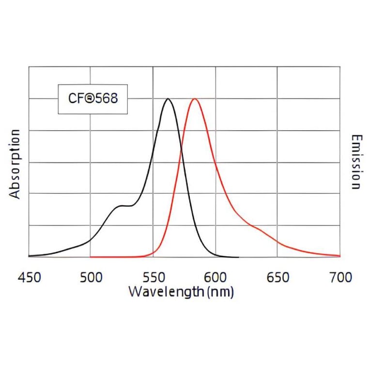
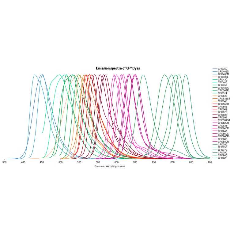
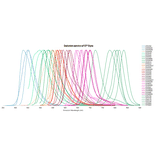
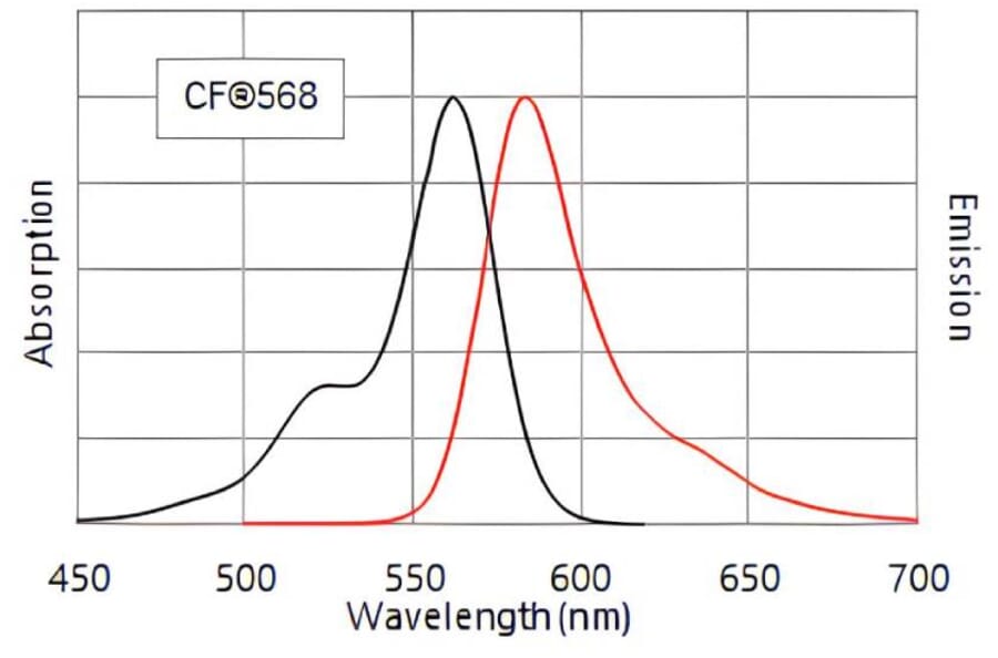
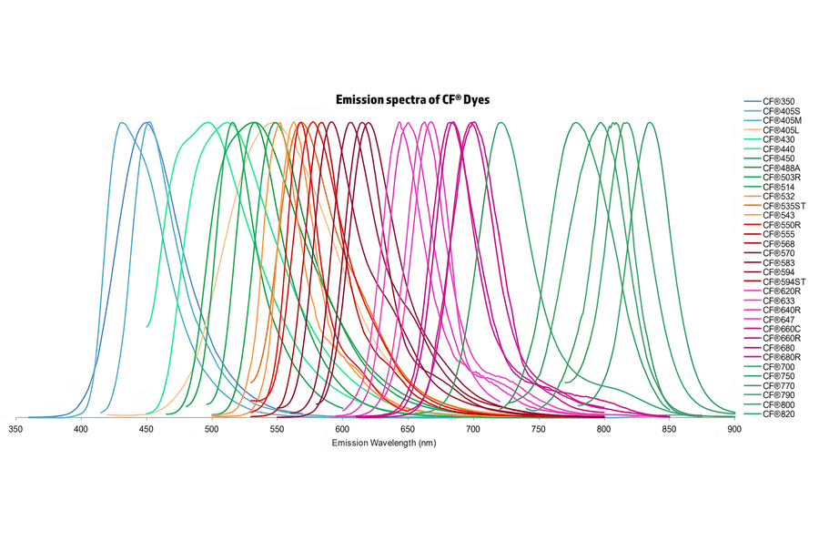
Normalized emission spectra of the CF® dye family spanning the visible to near-infrared range are shown, illustrating the spectral diversity and overlap between dyes. Curves represent relative fluorescence intensity as a function of emission wavelength (nm), with peak positions corresponding to each dye’s characteristic emission maximum. This reference highlights the broad coverage of CF® dyes for multicolor fluorescence applications and aids in selecting compatible dye combinations for imaging, flow cytometry, and other fluorescence-based assays.
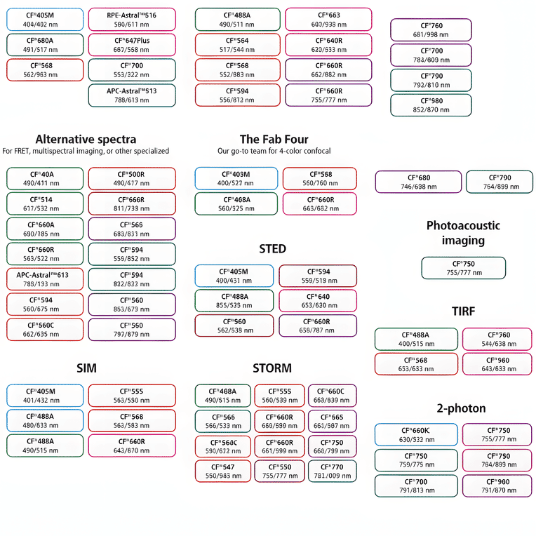
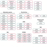
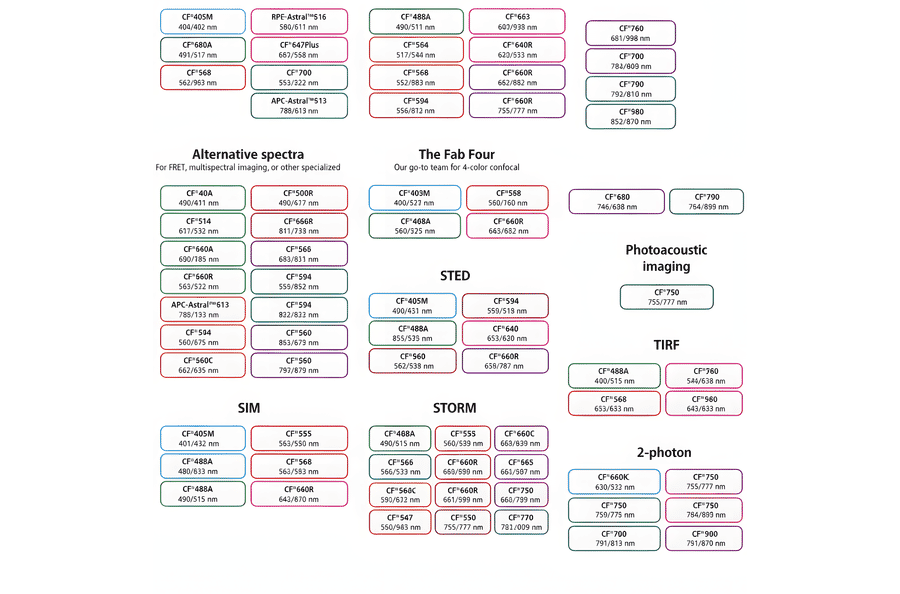
This chart summarizes commonly used CF® dyes grouped by their suitability for specific imaging modalities, including alternative spectra applications, four-color confocal imaging, near-infrared western blotting, photoacoustic imaging, STED, SIM, STORM, TIRF, and two-photon microscopy. Each dye is shown with its characteristic excitation and emission wavelengths (nm), providing a practical reference for selecting spectrally compatible dyes and optimizing multicolor experimental design across a range of fluorescence techniques.
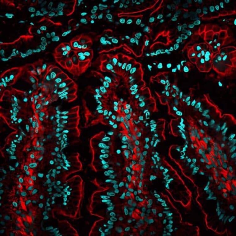
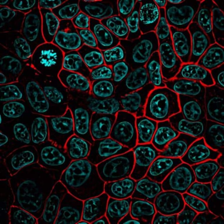
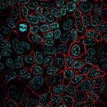
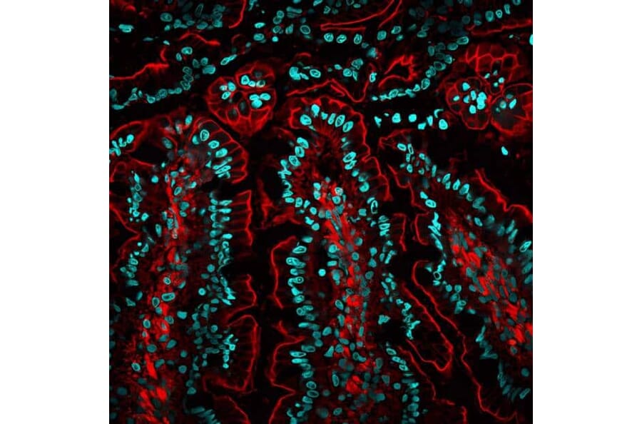
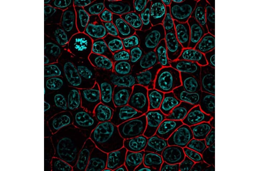
MCF-7 cells stained with a CF®568-conjugated monoclonal anti-Ep-CAM antibody (clone EGP40/826) to label Ep-CAM (red), with nuclei counterstained using Hoechst (blue).
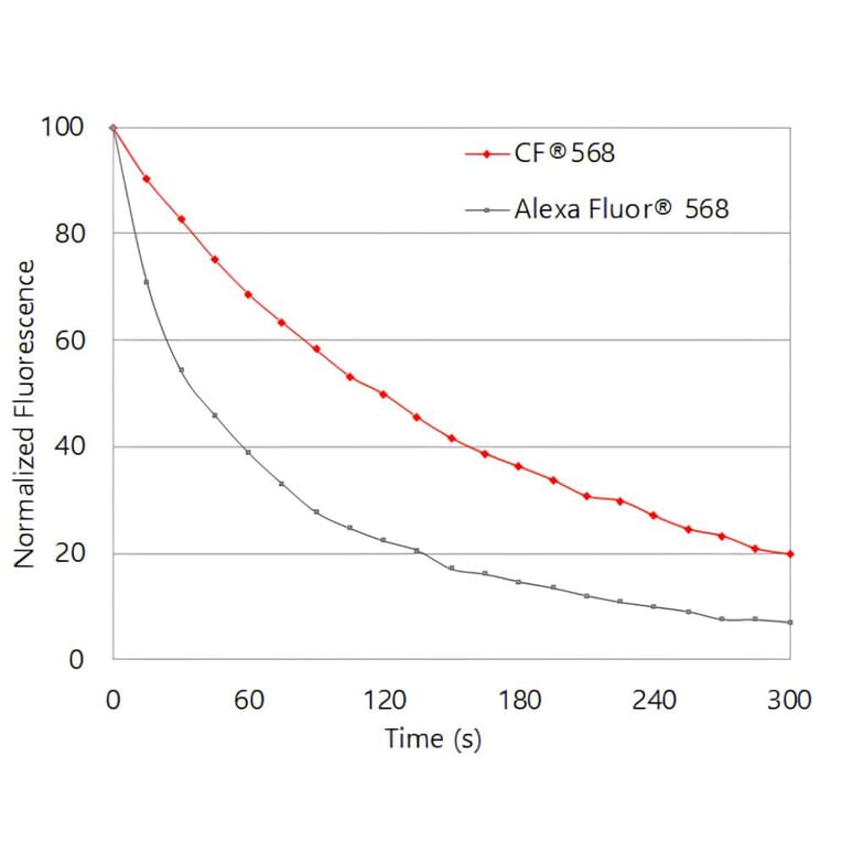
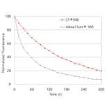
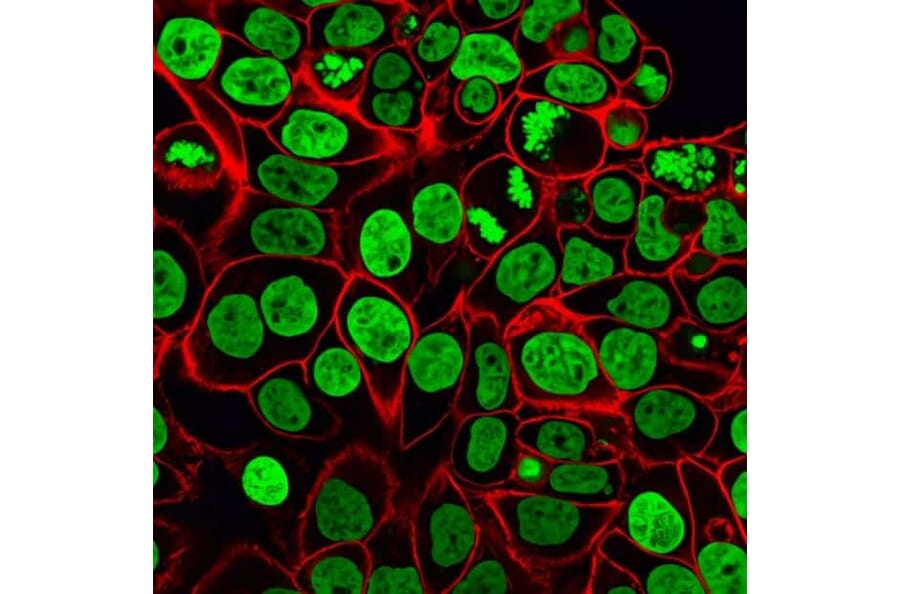
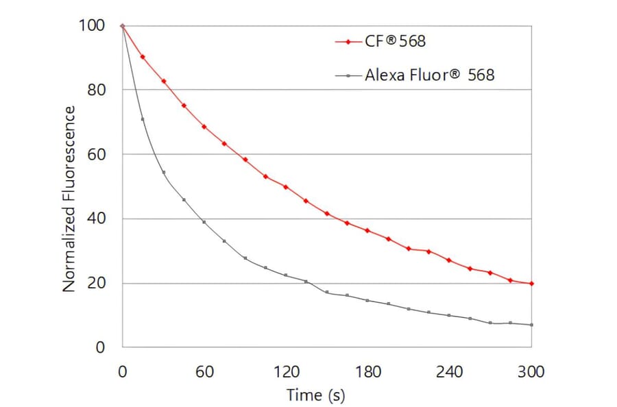
CF®568 exhibits greater photostability than Alexa Fluor® 568. Jurkat cells were stained with biotinylated anti-CD3 antibody followed by CF®568 or Alexa Fluor® 568 streptavidin conjugates. Cells were continuously illuminated using a mercury arc lamp with a Cy®3 filter cube. Images were acquired every 15 seconds for 5 minutes, and fluorescence intensity was normalized to the initial time point.