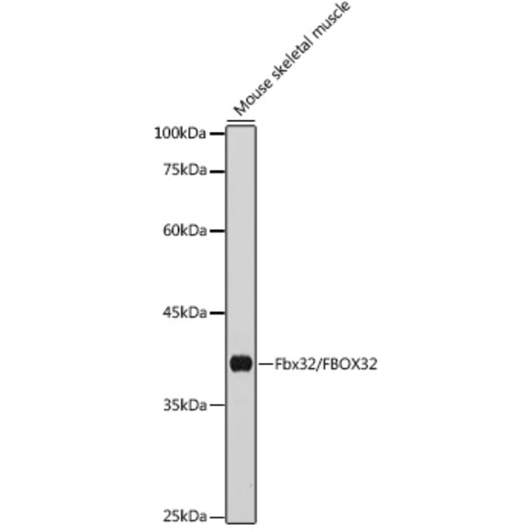
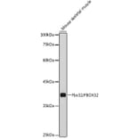
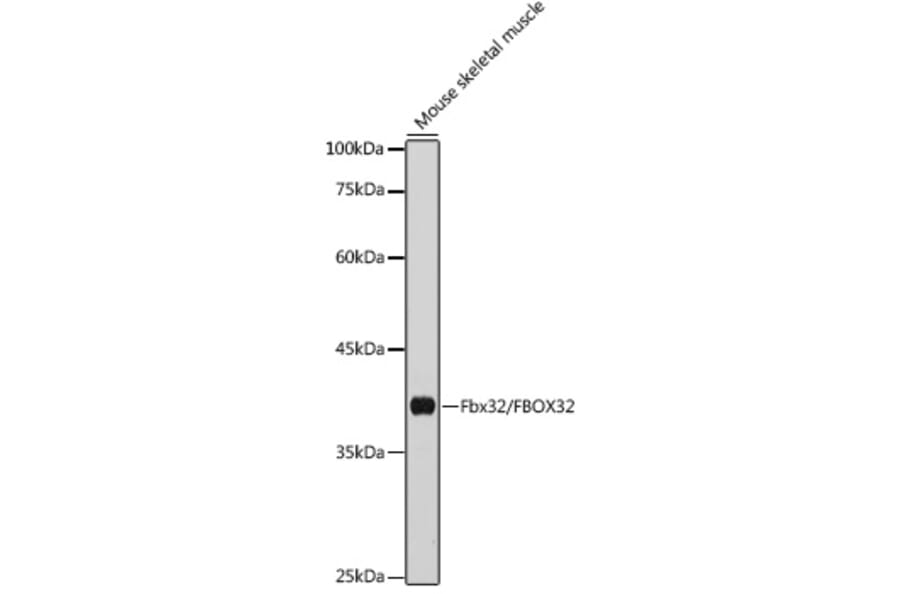
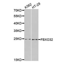
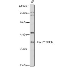
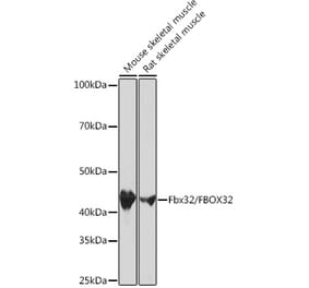
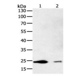
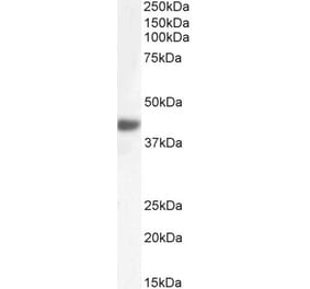
Western blot analysis of extracts of Mouse skeletal muscle, using Anti-Fbx32 Antibody (A14404) at 1:1,000 dilution. The secondary antibody was Goat Anti-Rabbit IgG H&L Antibody (HRP) at 1:10,000 dilution. Lysates/proteins were present at 25µg per lane. The blocking buffer used was 3% non-fat dry milk in TBST. Detection was with a ECL Basic Kit. Exposure time: 10s.
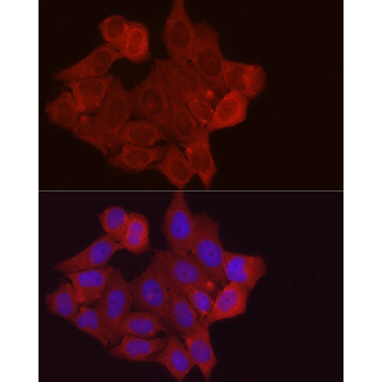
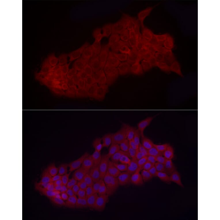
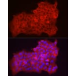
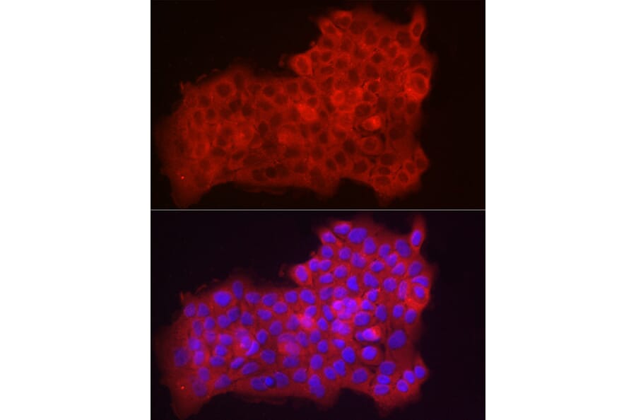
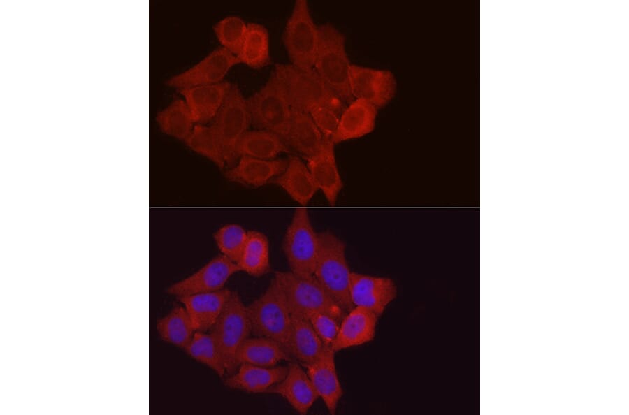
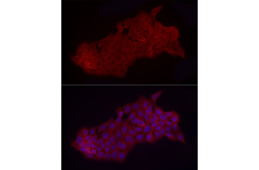
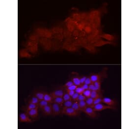
Immunofluorescence analysis of A431 cells using Anti-Fbx32 Antibody (A14404) at a dilution of 1:50 (40x lens). DAPI was used to stain the cell nuclei (blue).
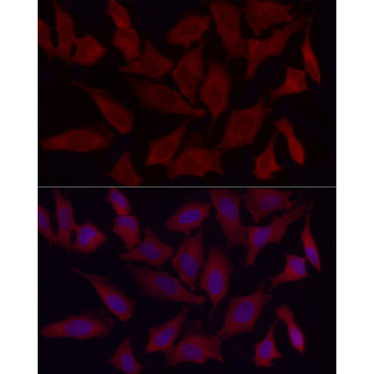
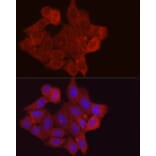
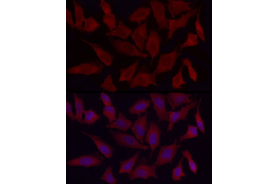
Immunofluorescence analysis of HeLa cells using Anti-Fbx32 Antibody (A14404) at a dilution of 1:50 (40x lens). DAPI was used to stain the cell nuclei (blue).
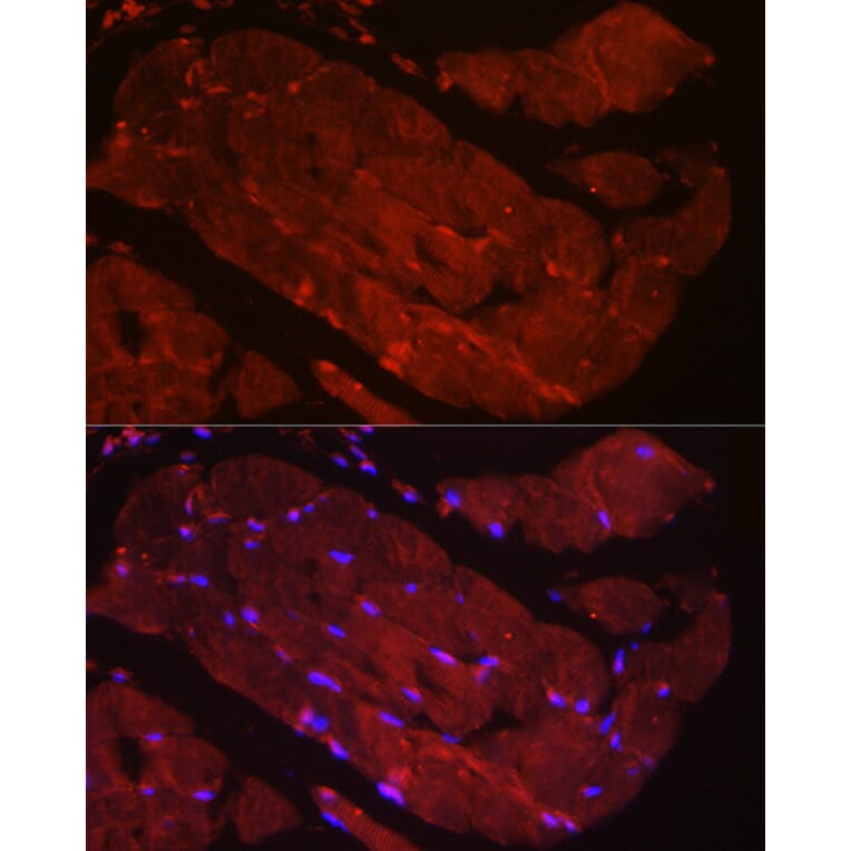
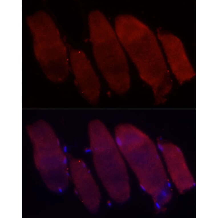
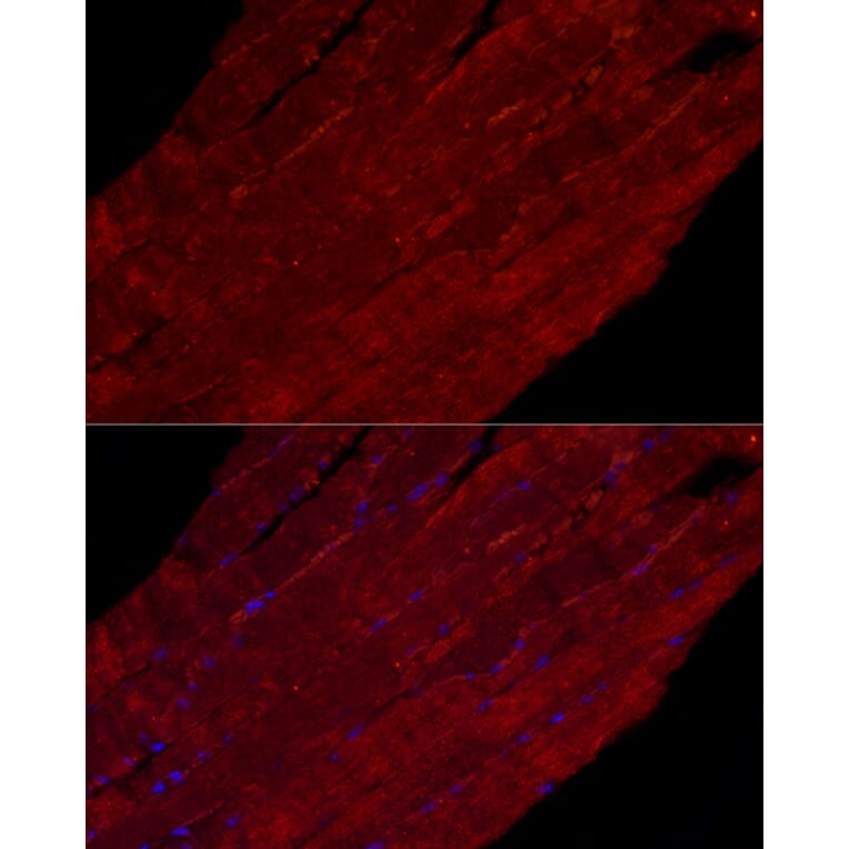
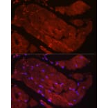
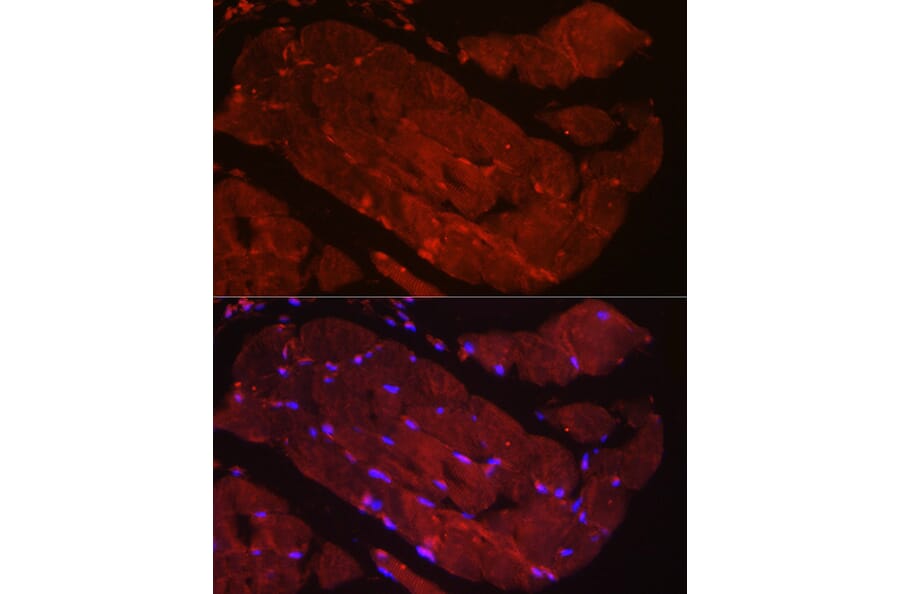
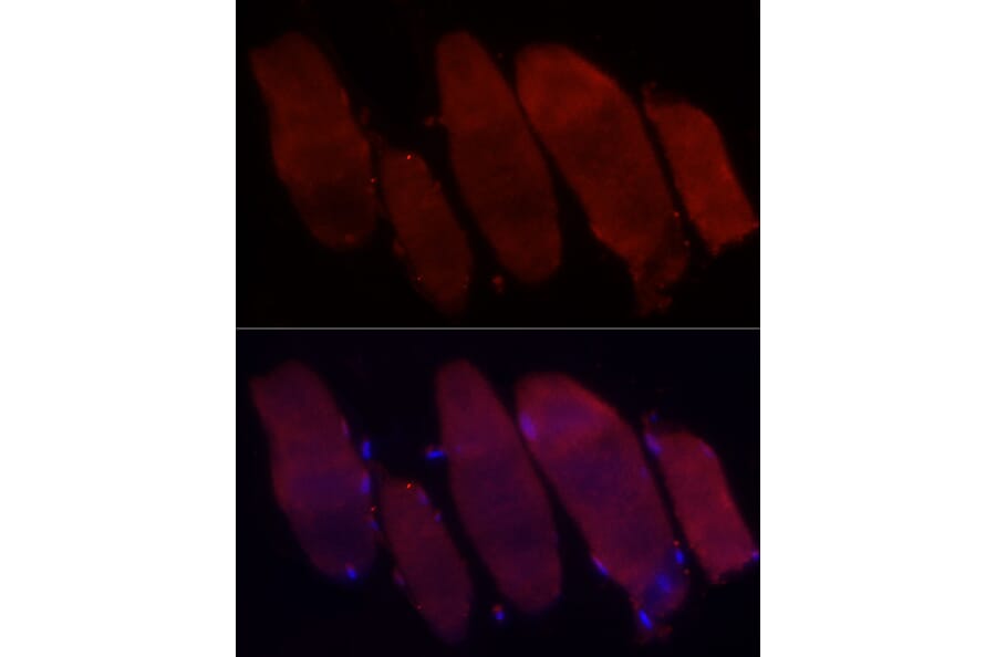
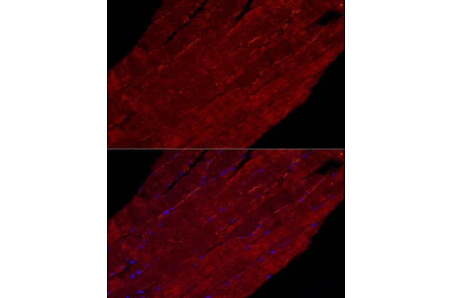
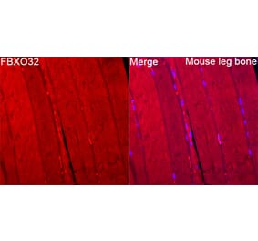
Immunofluorescence analysis of mouse skeletal muscle cells using Anti-Fbx32 Antibody (A14404) at a dilution of 1:50 (40x lens). DAPI was used to stain the cell nuclei (blue).
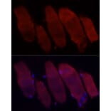
Immunofluorescence analysis of rat skeletal muscle cells using Anti-Fbx32 Antibody (A14404) at a dilution of 1:50 (40x lens). DAPI was used to stain the cell nuclei (blue).
Immunofluorescence analysis of A431 cells using Anti-Fbx32 Antibody (A14404) at a dilution of 1:50 (40x lens). DAPI was used to stain the cell nuclei (blue).
Immunofluorescence analysis of HeLa cells using Anti-Fbx32 Antibody (A14404) at a dilution of 1:50 (40x lens). DAPI was used to stain the cell nuclei (blue).
Immunofluorescence analysis of mouse skeletal muscle cells using Anti-Fbx32 Antibody (A14404) at a dilution of 1:50 (40x lens). DAPI was used to stain the cell nuclei (blue).