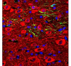
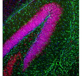
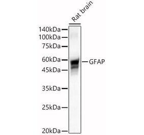
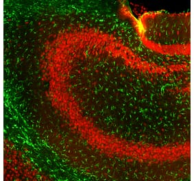
This antibody reacts with intact GFAP molecules. GFAP is the principal marker of astroglial cells in the central nervous system; it is specifically expressed in satellite cells in peripheral ganglia and in non myelinating Schwann cells in peripheral nerves. The GFAP protein runs on gels at ~55 kDa protein, usually associated with lower Mw bands which are thought to be proteolytic fragments and alternate transcripts from the single gene.
Applications
WB, IHC-P, IHC-Fr, ICC
Dilutions
WB: 1-2 µg/ml.
Reactivity
Human, Porcine
Immunogen
Pellet of porcine brain cold-stable proteins after depolymerization of microtubules.
Host
Mouse
Clonality
Monoclonal
Clone ID
GF-02
Isotype
IgM
Conjugate
Unconjugated
Purification
Purified by differential precipitation and solid-phase chromatography.
Concentration
1 mg/ml
Predicted MW
55 kDa
Product Form
Liquid
Formulation
Supplied in Tris Buffered Saline, pH 8, with 15 mM Sodium Azide.
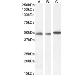
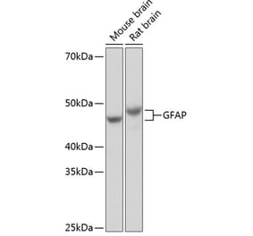
Western blotting analysis of human GFAP using Anti-GFAP Antibody [GF-02] (A85479) on lysates of Neuro 2a and HEK 293T cells under reducing and non-reducing conditions. Nitrocellulose membrane was probed with 2 µg/ml of mouse anti-GFAP monoclonal antibody followed by IRDye800-conjugated anti-mouse secondary antibody. A specific band was detected for GFAP at approximately 54 kDa.
Western blotting analysis (reducing conditions) of GFAP in porcine brain lysate. Lane 1: Immunostaining using Anti-GFAP Antibody [GF-02] (A85479)Lane 2: Immunostaining with Mouse IgM Isotype Control [PFR-03] (A86761).
Publishing research using Anti-GFAP Antibody [GF-02] (A85479)? Please let us know so that we can list the citation on this page.


![WB - Anti-GFAP Antibody [GF-02] (A85479)](https://cdn.antibodies.com/image/catalog/85/A85479_1.jpg?profile=product_top)
![WB - Anti-GFAP Antibody [GF-02] (A85479)](https://cdn.antibodies.com/image/catalog/85/A85479_2.jpg?profile=product_top)
![WB - Anti-GFAP Antibody [GF-02] (A85479)](https://cdn.antibodies.com/image/catalog/85/A85479_1.jpg?profile=product_top_thumb)
![WB - Anti-GFAP Antibody [GF-02] (A85479)](https://cdn.antibodies.com/image/catalog/85/A85479_2.jpg?profile=product_top_thumb)
![WB - Anti-GFAP Antibody [GF-02] (A85479)](https://cdn.antibodies.com/image/catalog/85/A85479_1.jpg?profile=product_image)
![WB - Anti-GFAP Antibody [GF-02] (A85479)](https://cdn.antibodies.com/image/catalog/85/A85479_2.jpg?profile=product_image)
![Immunofluorescence - Anti-GFAP Antibody [2A5] (A104314) - Antibodies.com](https://cdn.antibodies.com/image/catalog/104/A104314_1.jpg?profile=product_alternative)
![Immunofluorescence - Anti-GFAP Antibody [5C10] (A85422) - Antibodies.com](https://cdn.antibodies.com/image/catalog/85/A85422_1.jpg?profile=product_alternative)
![Western Blot - Anti-GFAP Antibody [GA-5] - BSA and Azide free (A251887) - Antibodies.com](https://cdn.antibodies.com/image/catalog/251/A251887_1.jpg?profile=product_alternative)
![Western Blot - Anti-GFAP Antibody [GA-5] (A248705) - Antibodies.com](https://cdn.antibodies.com/image/catalog/248/A248705_1.jpg?profile=product_alternative)
![Immunohistochemistry - Anti-GFAP Antibody [GA-5 + ASTRO/789] (A251890) - Antibodies.com](https://cdn.antibodies.com/image/catalog/248/A248709_1.jpg?profile=product_alternative)
![Immunohistochemistry - Anti-GFAP Antibody [SPM507] - BSA and Azide free (A248707) - Antibodies.com](https://cdn.antibodies.com/image/catalog/251/A251889_1.jpg?profile=product_alternative)
![Immunohistochemistry - Anti-GFAP Antibody [SPM248] - BSA and Azide free (A248706) - Antibodies.com](https://cdn.antibodies.com/image/catalog/251/A251888_1.jpg?profile=product_alternative)
![Immunohistochemistry - Anti-GFAP Antibody [SPM248] (A251891) - Antibodies.com](https://cdn.antibodies.com/image/catalog/248/A248706_1.jpg?profile=product_alternative)
![Immunohistochemistry - Anti-GFAP Antibody [GA-5 + ASTRO/789] - BSA and Azide free (A248709) - Antibodies.com](https://cdn.antibodies.com/image/catalog/251/A251891_1.jpg?profile=product_alternative)
![IHC - Anti-GFAP Antibody [GA-5] (A85508)](https://cdn.antibodies.com/image/catalog/85/A85508_1.jpg?profile=product_alternative)