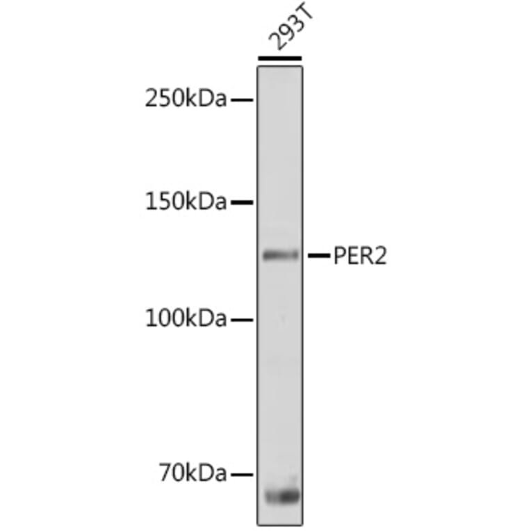
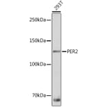
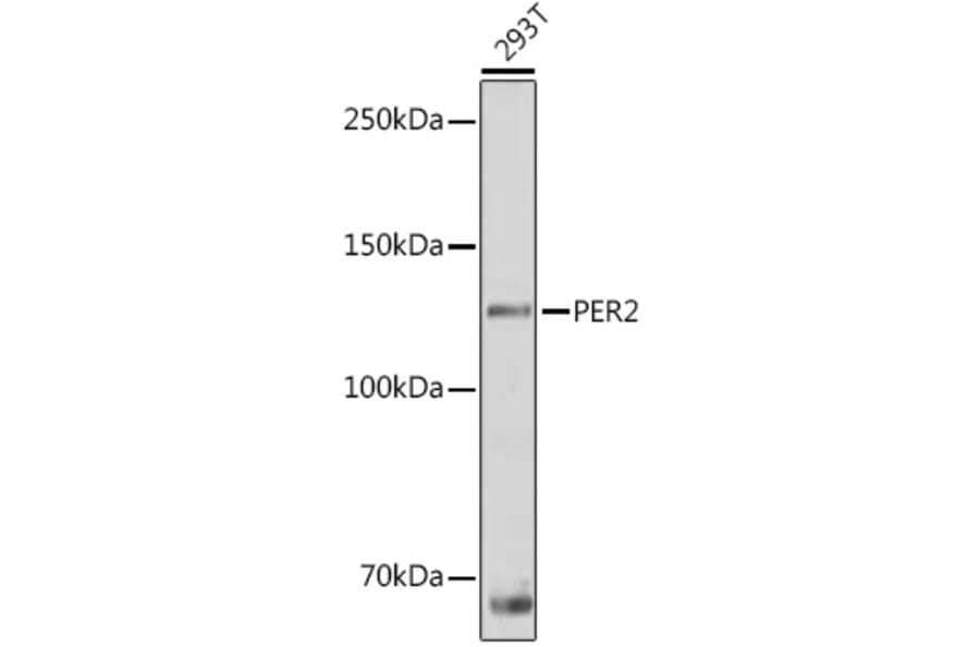
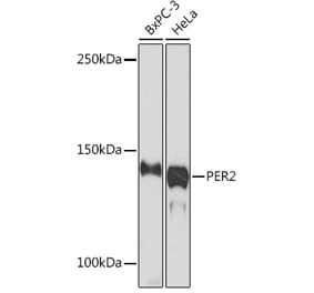
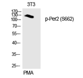
Western blot analysis of extracts of 293T cells, using Anti-PER2 Antibody (A88334) at 1:1,000 dilution. The secondary antibody was Goat Anti-Rabbit IgG H&L Antibody (HRP) at 1:10,000 dilution. Lysates/proteins were present at 25µg per lane. The blocking buffer used was 3% non-fat dry milk in TBST. Detection was with a ECL Basic Kit. Exposure time: 10s.
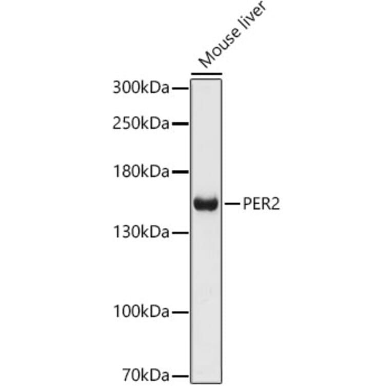
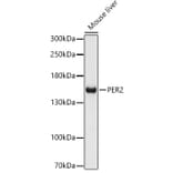
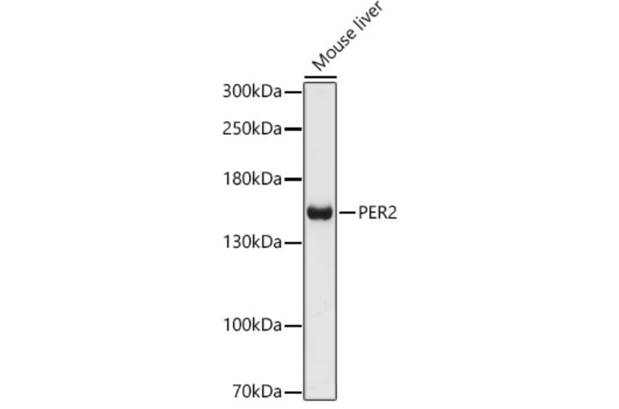
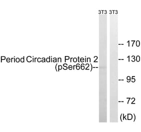
Western blot analysis of extracts of Mouse liver, using Anti-PER2 Antibody (A88334) at 1:1,000 dilution. The secondary antibody was Goat Anti-Rabbit IgG H&L Antibody (HRP) at 1:10,000 dilution. Lysates/proteins were present at 25µg per lane. The blocking buffer used was 3% non-fat dry milk in TBST. Detection was with a ECL Basic Kit. Exposure time: 90s.
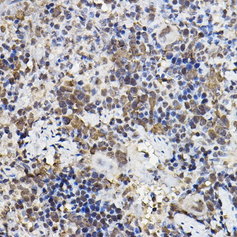
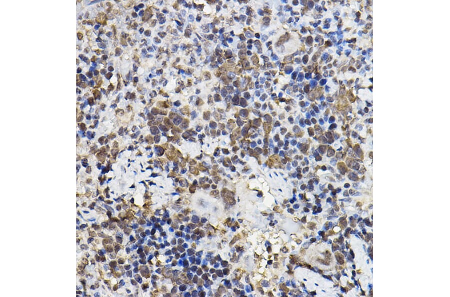
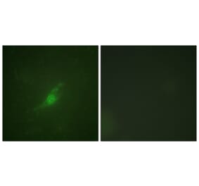
Immunohistochemistry analysis of paraffin-embedded rat spleen using Anti-PER2 Antibody (A88334) at a dilution of 1:100 (40x lens). Perform high pressure antigen retrieval with 10 mM citrate buffer pH 6.0 before commencing with IHC staining protocol.
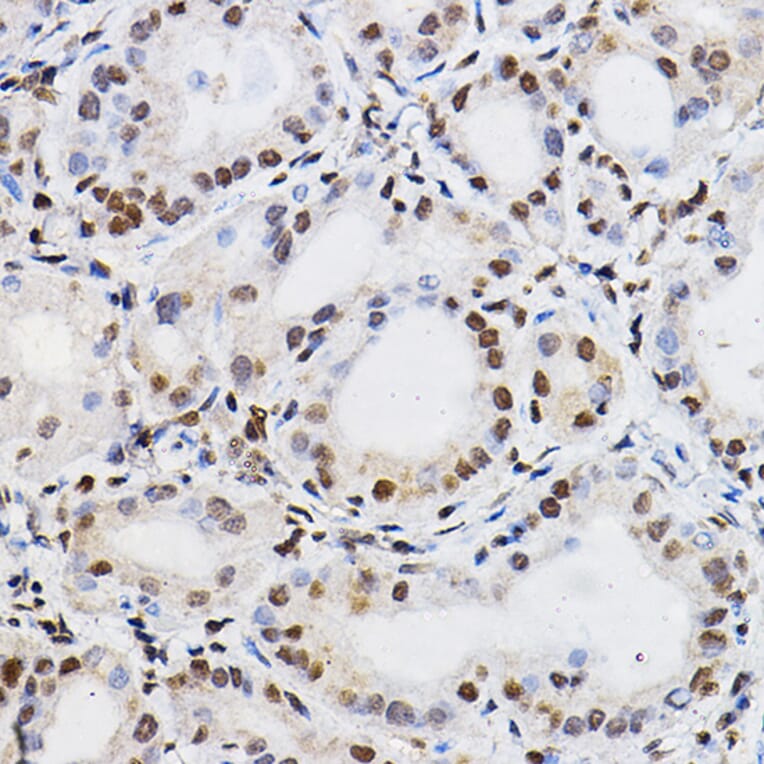
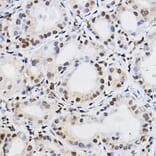
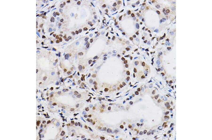
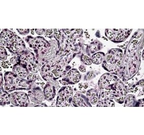
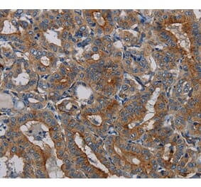
Immunohistochemistry analysis of paraffin-embedded human thyroid cancer using Anti-PER2 Antibody (A88334) at a dilution of 1:100 (40x lens). Perform high pressure antigen retrieval with 10 mM citrate buffer pH 6.0 before commencing with IHC staining protocol.
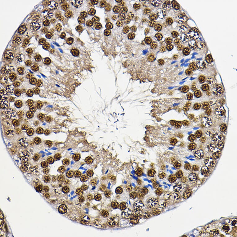
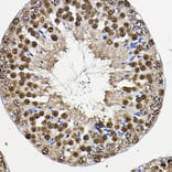
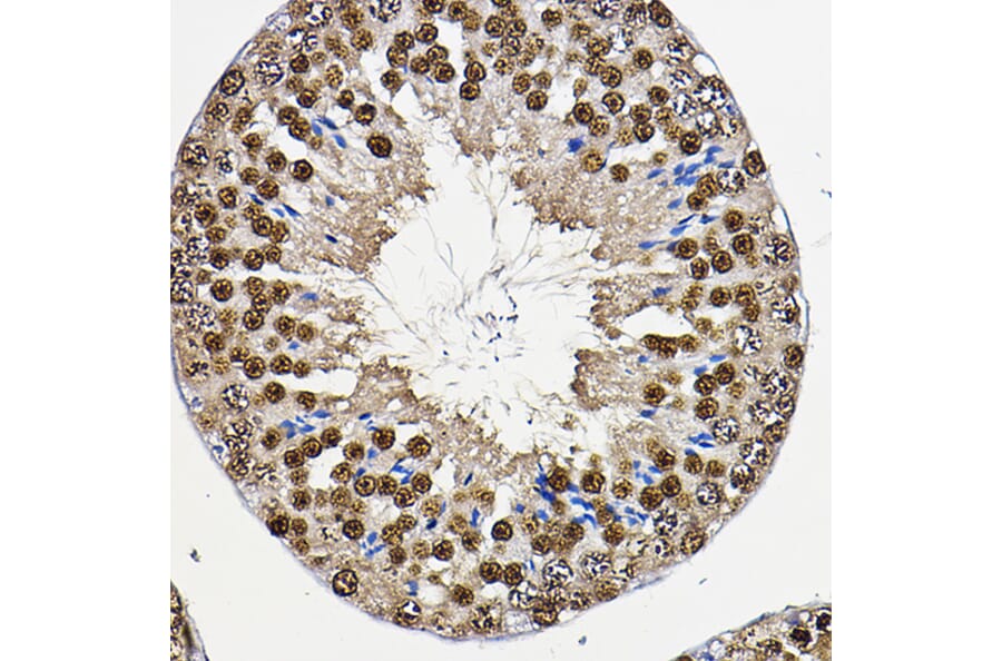
Immunohistochemistry analysis of paraffin-embedded mouse testis using Anti-PER2 Antibody (A88334) at a dilution of 1:100 (40x lens). Perform high pressure antigen retrieval with 10 mM citrate buffer pH 6.0 before commencing with IHC staining protocol.
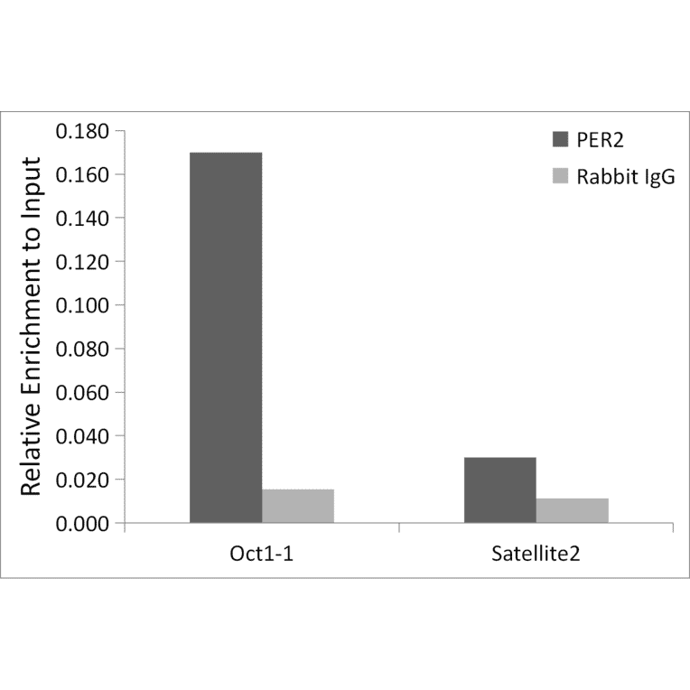
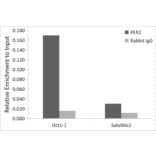
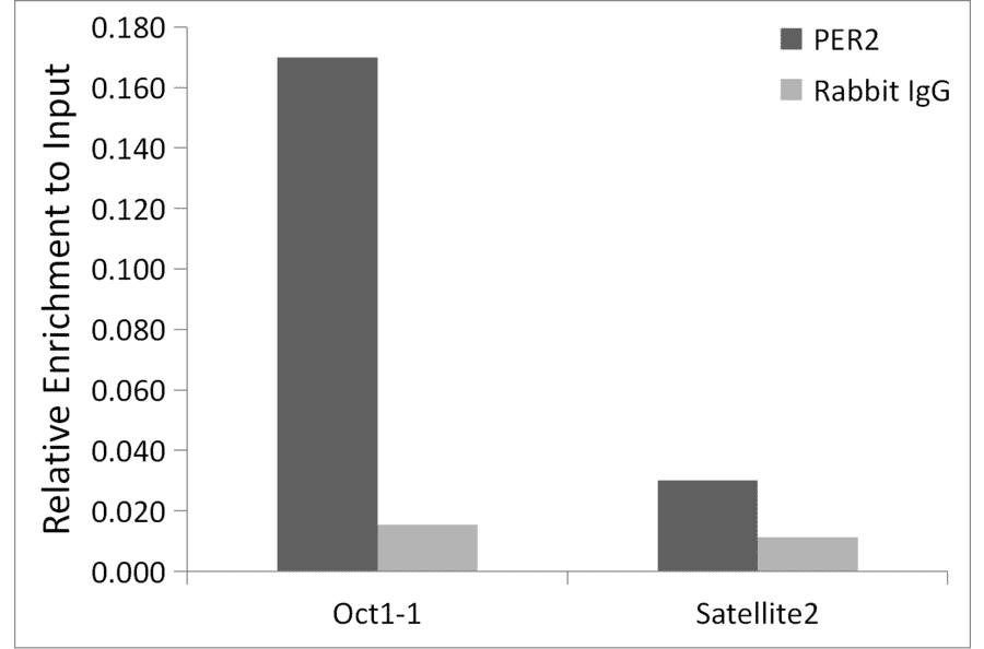
Chromatin immunoprecipitation (ChIP) analysis of extracts of MCF7 cells, using Anti-PER2 Antibody (A88334) and Rabbit IgG. The amount of immunoprecipitated DNA was checked by quantitative PCR. Histogram was constructed by the ratios of the immunoprecipitated DNA to the input.