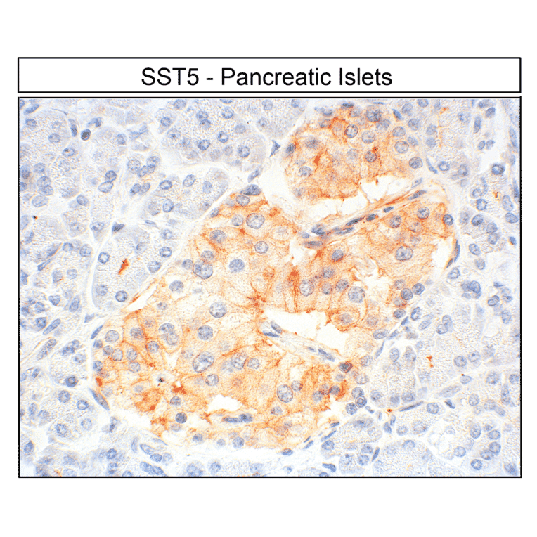
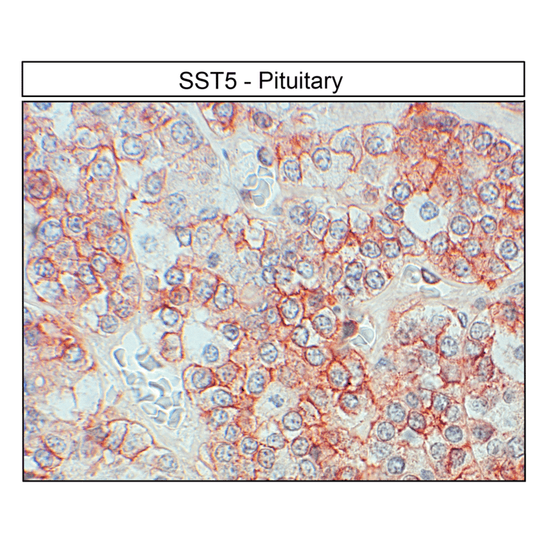
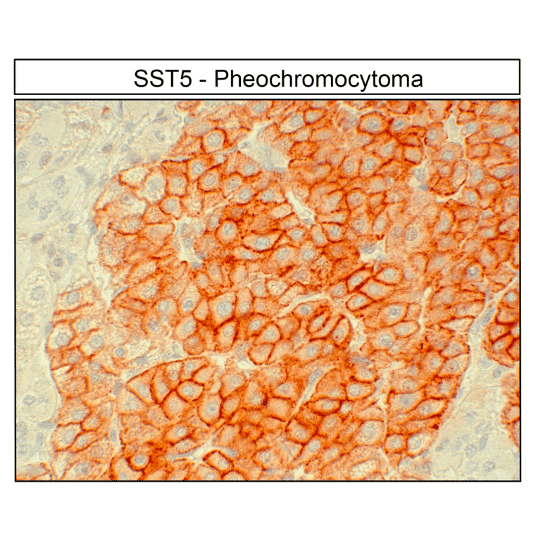
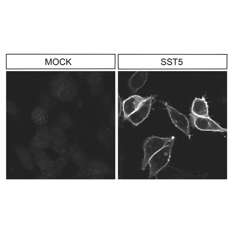
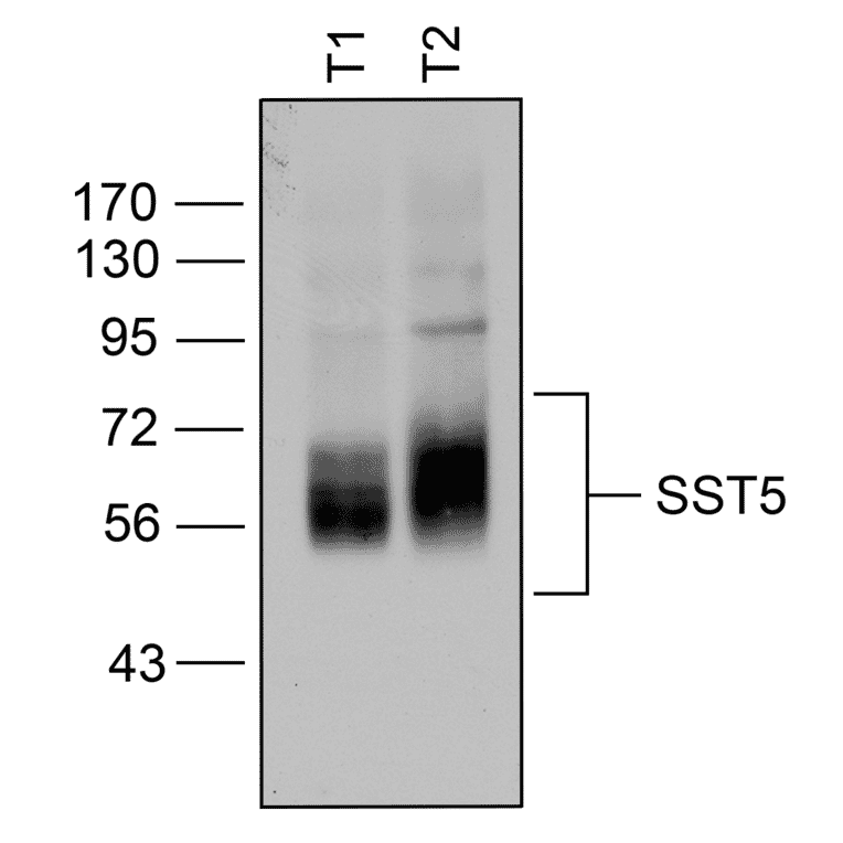
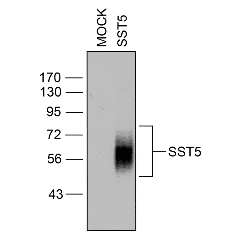
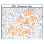
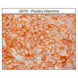
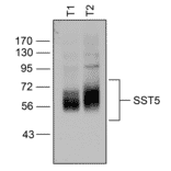
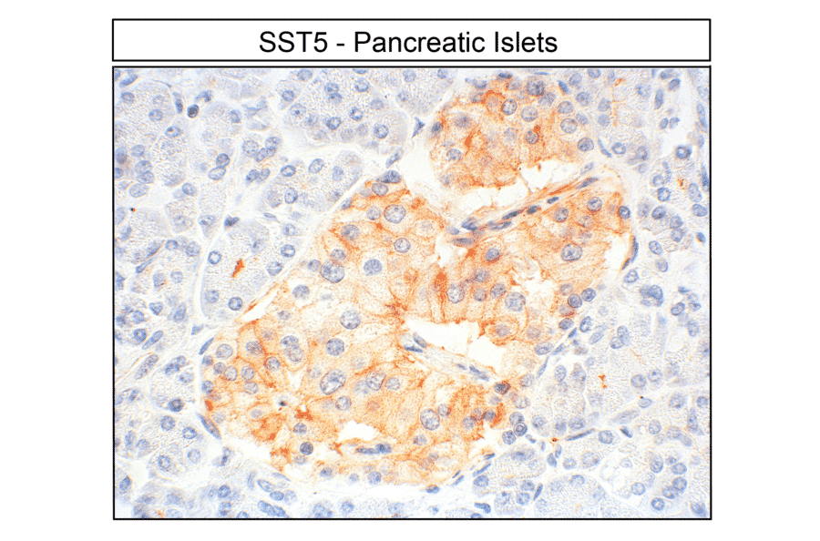
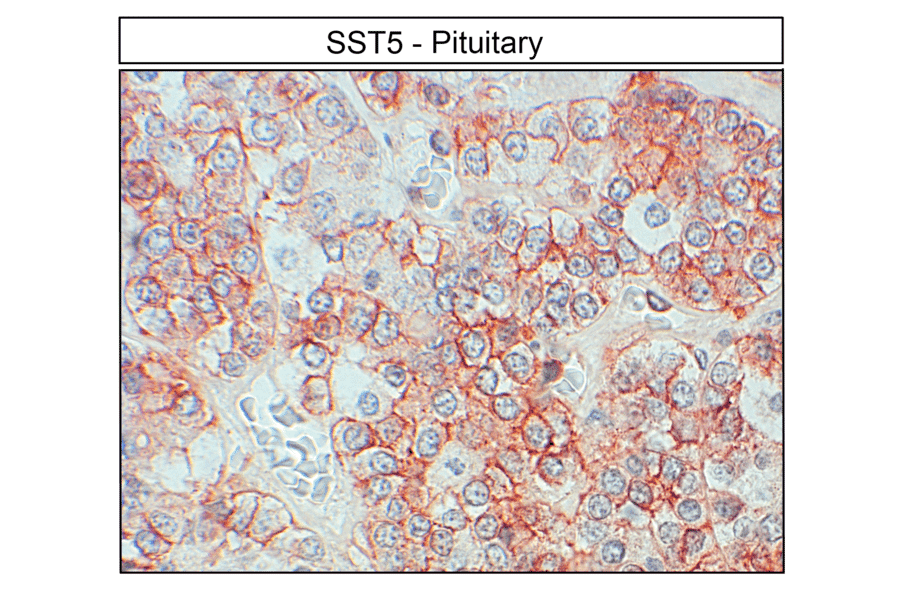
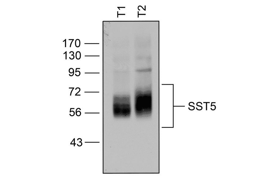
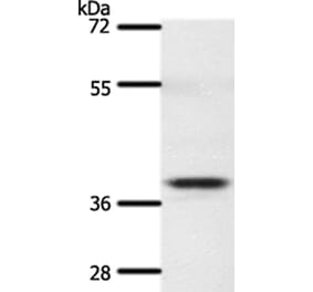
This antibody specifically recognizes the C-terminal region of human SST5, detecting the non-phosphorylated form. It is optimized for Western blot analysis, enables immunoprecipitation from cell and tissue lysates, and is suitable for immunohistochemistry in cultured cells and tissue sections. It is validated using siRNA-mediated knockdown (KO-Validated).
Applications
WB, ICC, IHC
Dilutions
WB: 1,000, IHC: 1:100, ICC: 1:200
Reactivity
Human
Immunogen
Synthetic peptide corresponding to human SST5 (amino acids. 344-364). Immunogen range is 22-22 amino acids.
Sequence
QEATPPAHRAAANGLMQTSKL
Host
Rabbit
Clonality
Polyclonal
Isotype
IgG
Conjugate
Unconjugated
Purification
Antigen affinity purification.
Concentration
Lot Specific
Product Form
Liquid
Formulation
Supplied in Dulbecco's PBS, pH 7.4, with 150 mM NaCl and 0.005% Sodium Azide.
Storage
Shipped at 4°C. Upon delivery aliquot and store at -20°C. Avoid freeze/thaw cycles.
Synonyms
Somatostatin receptor type 5, SS-5-R, SS5-R, SS5R, SST5
Immunohistochemical detection of Somatostatin Receptor 5 (SST5) in human pancreatic islets. Pancreatic sections were dewaxed, microwaved in citric acid, and stained with anti-SST5 antibody (A334481) at 1:100, followed by biotinylated anti-rabbit IgG and avidin-biotin solution. Color was developed with 3-amino-9-ethylcarbazole (AEC), and sections were counterstained with hematoxylin. SST5 receptors were detected at the plasma membrane of nearly all pancreatic islet cells.
Localization of Somatostatin Receptor 5 (SST5) in human anterior pituitary by immunohistochemistry. Anterior pituitary sections were dewaxed, microwaved in citric acid, and incubated with anti-SST5 antibody (A334481) at 1:100, followed by biotinylated anti-rabbit IgG and avidin-biotin solution. Color was developed with 3-amino-9-ethylcarbazole (AEC), and sections were counterstained with hematoxylin. SST5 receptors were observed in a distinct population of anterior pituitary cells.
Immunohistochemical detection of Somatostatin Receptor 5 (SST5) in human pheochromocytoma. Tumor sections were dewaxed, microwaved in citric acid, and stained with anti-SST5 antibody (A334481) at 1:100, followed by biotinylated anti-rabbit IgG and avidin-biotin solution. Color was developed with 3-amino-9-ethylcarbazole (AEC), and sections were counterstained with hematoxylin. SST5 receptors were uniformly detected at the plasma membrane of nearly all pheochromocytoma cells.
Localization of Somatostatin Receptor 5 (SST5) in human growth-hormone-producing pituitary adenoma by immunohistochemistry. Tumor sections were dewaxed, microwaved in citric acid, and incubated with anti-SST5 antibody (A334481) at 1:100, followed by biotinylated anti-rabbit IgG and avidin-biotin solution. Sections were developed with 3,3-diaminobenzidine (DAB)-glucose oxidase and lightly counterstained with hematoxylin. SST5 receptors were uniformly detected at the plasma membrane of nearly all tumor cells.
Immunocytochemical analysis of Somatostatin Receptor 5 (SST5) in transfected HEK293 cells. Native HEK293 cells (MOCK) or cells stably expressing SST5 were stained with anti-SST5 antibody (A334481) at 1:200. SST5 receptors were observed at the plasma membrane in transfected cells.
Western blot analysis of Somatostatin Receptor 5 (SST5) in human neuroendocrine tumors. Homogenates from neuroendocrine tumor samples (T1, T2) were immunoblotted with anti-SST5 antibody (A334481) at 1:1000.
Western blot confirmation of Somatostatin Receptor 5 (SST5) in transfected HEK293 cells. Lysates from native HEK293 cells (MOCK) or cells stably expressing SST5 were probed with anti-SST5 antibody (A334481) at 1:1000.