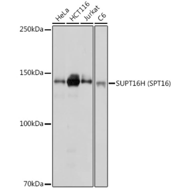
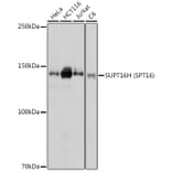
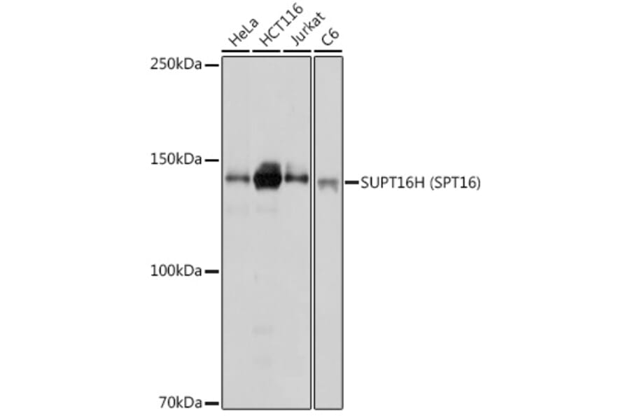
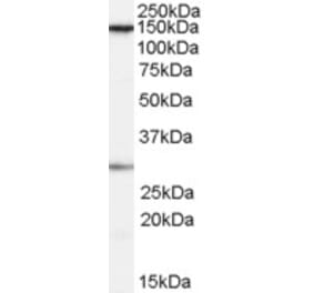
Western blot analysis of extracts of various cell lines, using Anti-SUPT16H Antibody (A306383) at 1:1,000 dilution. The secondary antibody was Goat Anti-Rabbit IgG H&L Antibody (HRP) at 1:10,000 dilution. Lysates/proteins were present at 25µg per lane. The blocking buffer used was 3% non-fat dry milk in TBST. Detection was with a ECL Basic Kit. Exposure time: 1s.
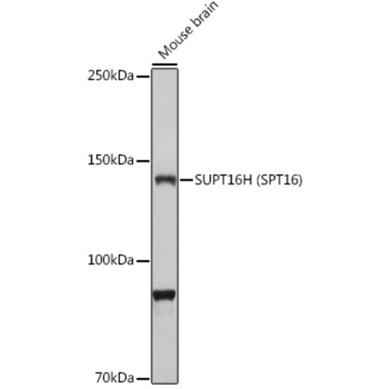
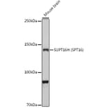
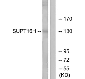
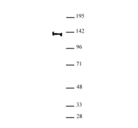
Western blot analysis of extracts of Mouse brain, using Anti-SUPT16H Antibody (A306383) at 1:1,000 dilution. The secondary antibody was Goat Anti-Rabbit IgG H&L Antibody (HRP) at 1:10,000 dilution. Lysates/proteins were present at 25µg per lane. The blocking buffer used was 3% non-fat dry milk in TBST. Detection was with a ECL Basic Kit. Exposure time: 30s.
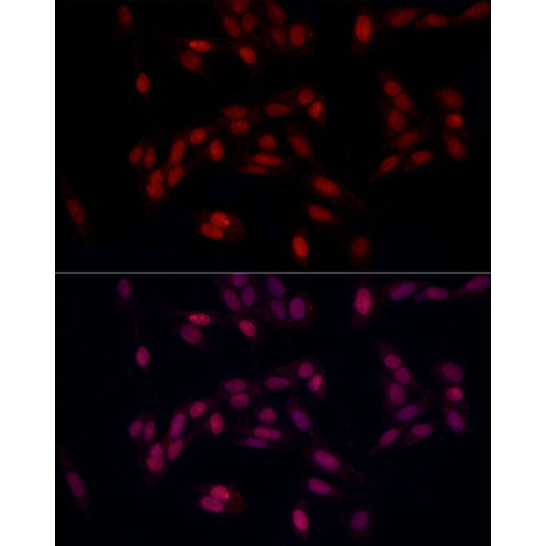
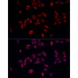
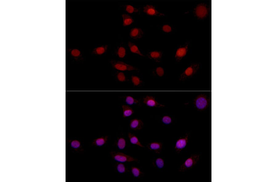
Immunofluorescence analysis of U2OS using Anti-SUPT16H Antibody (A306383) at a dilution of 1:100 (40x lens). DAPI was used to stain the cell nuclei (blue).
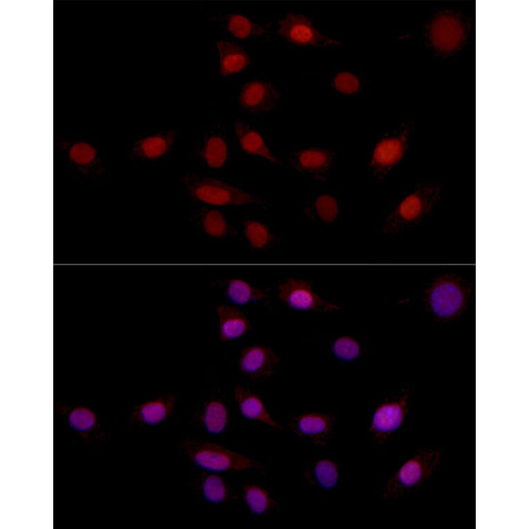
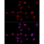
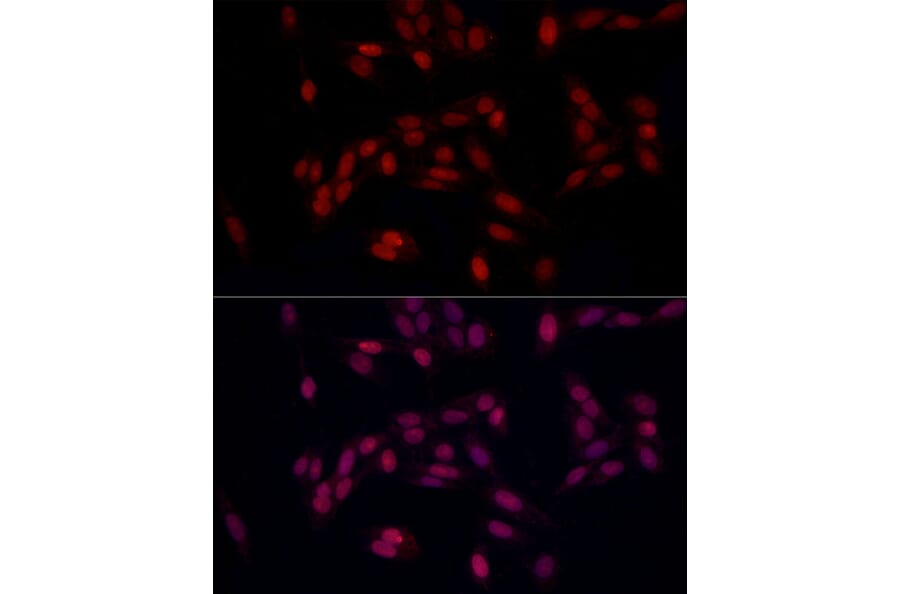
Immunofluorescence analysis of NIH/3T3 using Anti-SUPT16H Antibody (A306383) at a dilution of 1:100 (40x lens). DAPI was used to stain the cell nuclei (blue).
Publishing research using Anti-SUPT16H Antibody (A306383)? Please let us know so that we can list the citation on this page.
Alternative products to Anti-SUPT16H Antibody (A306383)