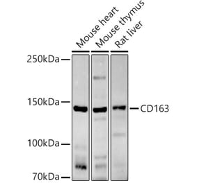
This antibody recognizes CD163, an approximately 130 kDa high affinity scavenger receptor expressed mainly on monocytes and macrophages, which binds hemoglobin-haptoglobin complex.
Applications
Flow Cytometry, IP, WB, IHC-Fr
Dilutions
Flow Cytometry: 1-4 µg/ml
Reactivity
Human
Immunogen
Hairy cell leukemia cells.
Host
Mouse
Clonality
Monoclonal
Clone ID
GHI/61
Isotype
IgG1
Light Chains
kappa
Conjugate
Unconjugated
Purification
Protein A chromatography.
Concentration
1 mg/ml
Predicted MW
130 kDa
Product Form
Liquid
Formulation
Supplied in Phosphate Buffered Saline, pH 7.4, with 15 mM Sodium Azide.
Storage
Shipped at 4°C. Upon delivery aliquot and store at -20°C. Avoid freeze/thaw cycles.
Synonyms
Hemoglobin scavenger receptor, M130, Scavenger receptor cysteine-rich type 1 protein M130
Separation of human monocytes (red-filled) from lymphocytes (black-dashed) in flow cytometry surface staining of human peripheral whole blood, labeling CD163 with Anti-CD163 Antibody [GHI/61] (A86219), (concentration in sample 2 µg/ml) GAM APC.
Publishing research using Anti-CD163 Antibody [GHI/61] (A86219)? Please let us know so that we can list the citation on this page.
Alternative products to Anti-CD163 Antibody [GHI/61] (A86219)


![Flow Cytometry - Anti-CD163 Antibody [GHI/61] (A86219)](https://cdn.antibodies.com/image/catalog/86/A86219_1.jpg?profile=product_top)
![Flow Cytometry - Anti-CD163 Antibody [GHI/61] (A86219)](https://cdn.antibodies.com/image/catalog/86/A86219_2.jpg?profile=product_top)
![Flow Cytometry - Anti-CD163 Antibody [GHI/61] (A86219)](https://cdn.antibodies.com/image/catalog/86/A86219_1.jpg?profile=product_top_thumb)
![Flow Cytometry - Anti-CD163 Antibody [GHI/61] (A86219)](https://cdn.antibodies.com/image/catalog/86/A86219_2.jpg?profile=product_top_thumb)
![Flow Cytometry - Anti-CD163 Antibody [GHI/61] (A86219)](https://cdn.antibodies.com/image/catalog/86/A86219_1.jpg?profile=product_image)
![Flow Cytometry - Anti-CD163 Antibody [GHI/61] (A86219)](https://cdn.antibodies.com/image/catalog/86/A86219_2.jpg?profile=product_image)
![Immunofluorescence - Anti-CD163 Antibody [EDHu-1] (A280344) - Antibodies.com](https://cdn.antibodies.com/image/catalog/280/A280344_14.jpg?profile=product_alternative)
![Immunohistochemistry - Anti-CD163 Antibody [ED2] (A280328) - Antibodies.com](https://cdn.antibodies.com/image/catalog/280/A280328_1.jpg?profile=product_alternative)
![Immunohistochemistry - Anti-CD163 Antibody [M130/1210] (A253762) - Antibodies.com](https://cdn.antibodies.com/image/catalog/250/A250603_1.jpg?profile=product_alternative)
![Immunohistochemistry - Anti-CD163 Antibody [M130/2163] - BSA and Azide free (A253785) - Antibodies.com](https://cdn.antibodies.com/image/catalog/253/A253785_1.jpg?profile=product_alternative)
![Immunohistochemistry - Anti-CD163 Antibody [M130/2162] - BSA and Azide free (A250604) - Antibodies.com](https://cdn.antibodies.com/image/catalog/253/A253784_1.jpg?profile=product_alternative)
![Immunohistochemistry - Anti-CD163 Antibody [M130/1210] - BSA and Azide free (A250603) - Antibodies.com](https://cdn.antibodies.com/image/catalog/253/A253783_1.jpg?profile=product_alternative)
![Immunohistochemistry - Anti-CD163 Antibody [M130/2164] - BSA and Azide free (A250606) - Antibodies.com](https://cdn.antibodies.com/image/catalog/253/A253786_1.jpg?profile=product_alternative)
![Immunohistochemistry - Anti-CD163 Antibody [M130/2164] (A253784) - Antibodies.com](https://cdn.antibodies.com/image/catalog/250/A250606_1.jpg?profile=product_alternative)