

Unconjugated
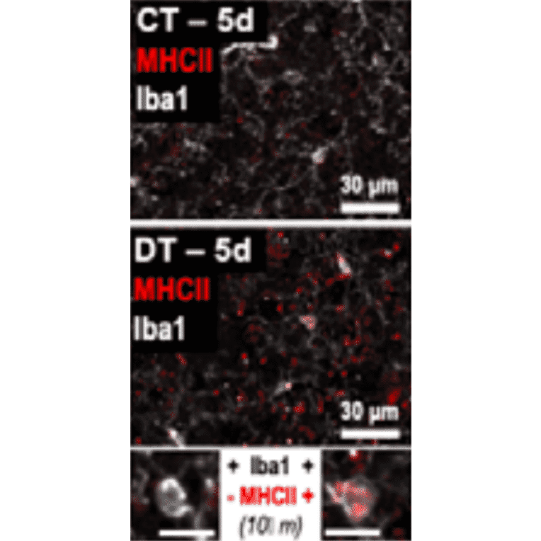
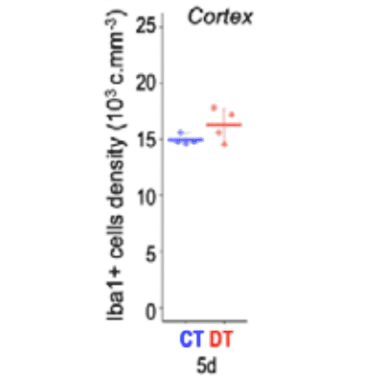
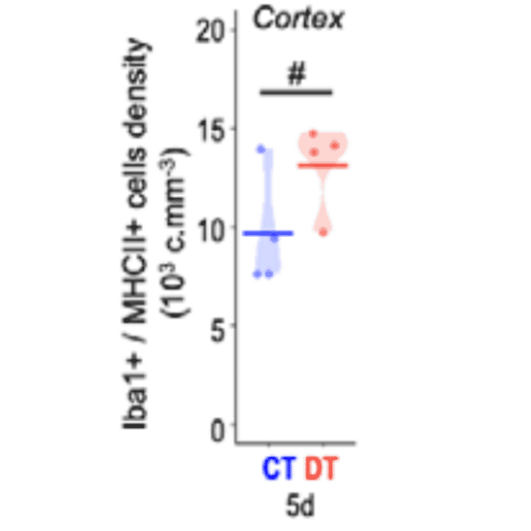
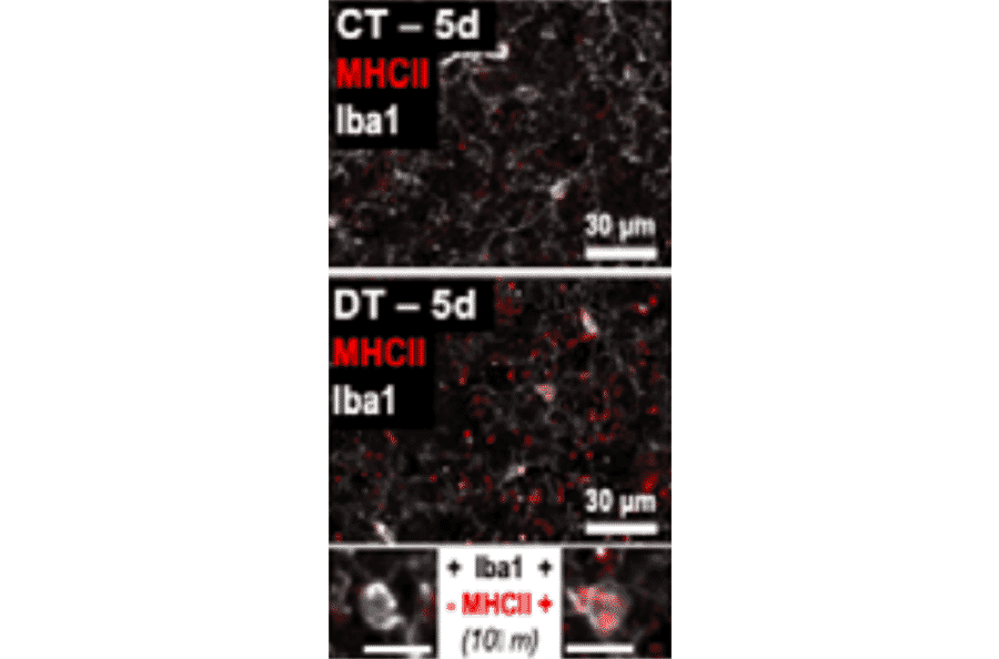
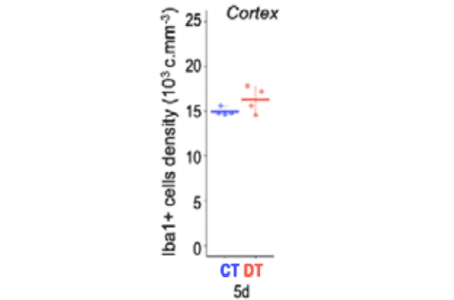
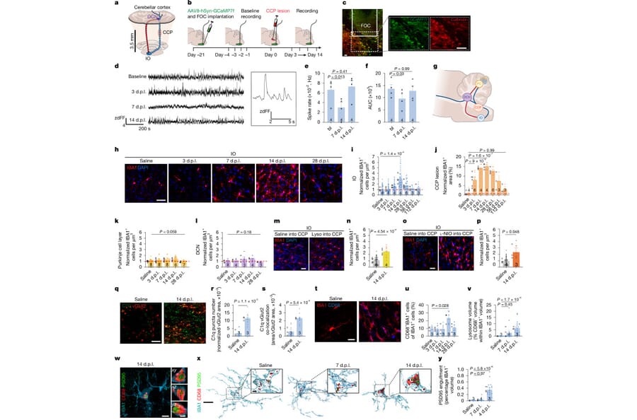
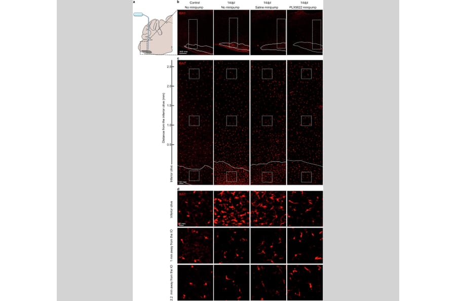
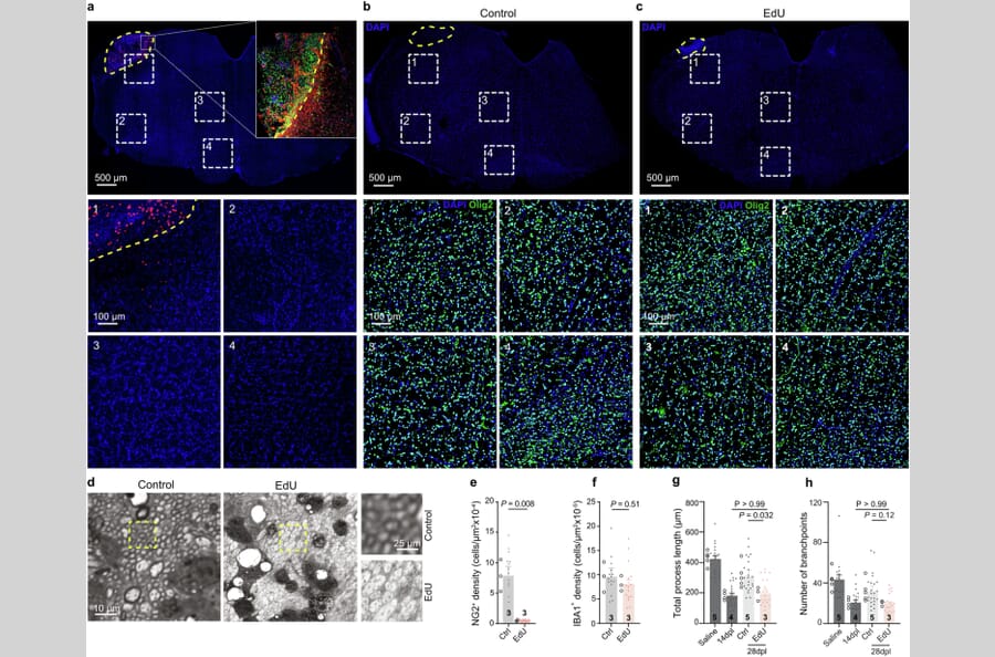
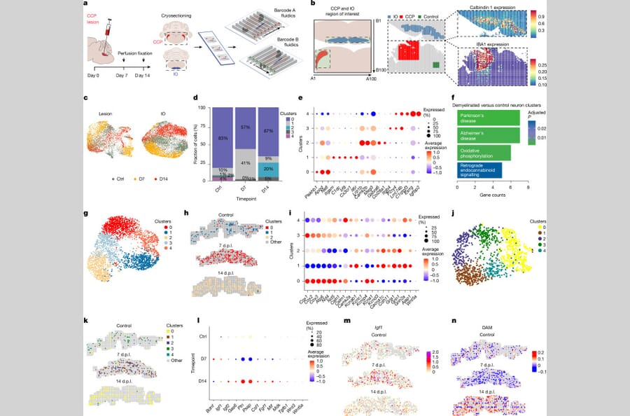
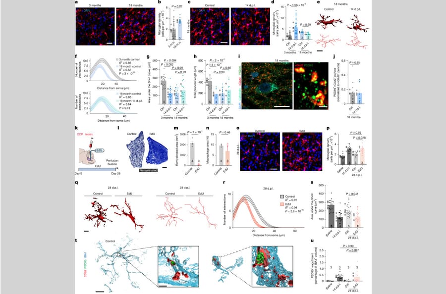
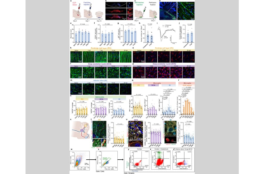
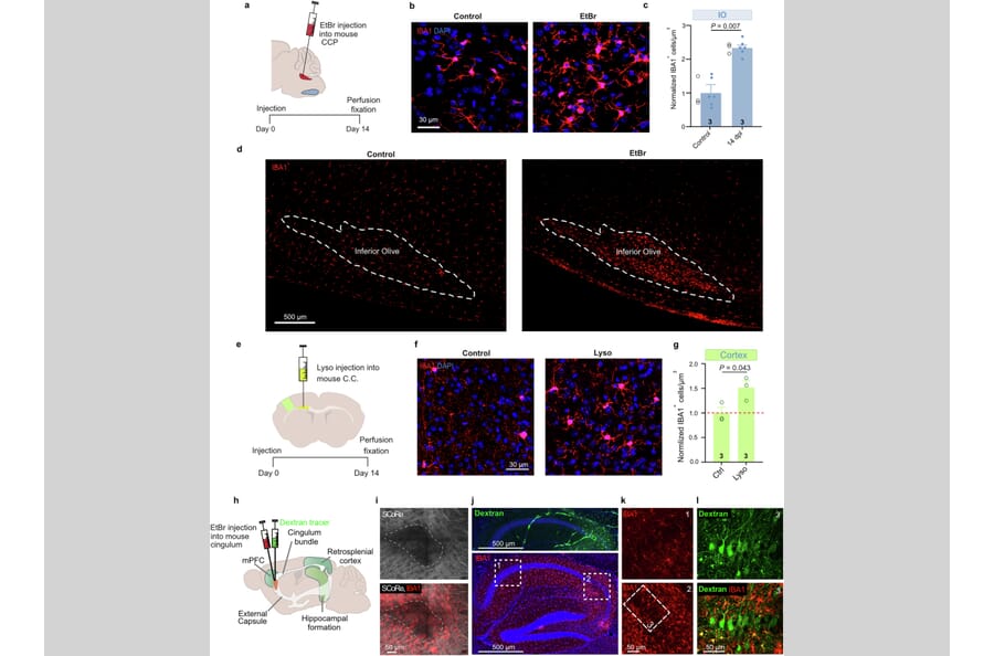
Focal white matter lesions occur in most neurodegenerative disorders1-3. Despite occurring early in disease, white matter lesions are considered to be independent of, or secondary to, grey matter neuroinflammation, synapse loss and altered neuronal activity4-7. Notably, their functional effect on neuronal circuits remains understudied. To address this, we generated a focal white matter lesion in the rat brain within a clinically relevant, anatomically well-defined circuit, in which these lesions occur in many neurodegenerative disorders8-10. Here we show that focal white matter lesions evoke transient neuronal activity changes and microgliosis, with subsequent synapse loss and increased microglial engulfment in the grey matter, which is reversed if myelin regeneration completes. Grey matter microgliosis is often considered to be detrimental; however, we show that it is an integral part of regeneration and is conserved across three distinct mouse circuits and lesioning methods. Preventing these transient changes in the grey matter blocks myelin regeneration in the white matter. Conversely, inducing myelin regeneration failure leads to chronic grey matter neuroinflammation. This recapitulates the low-grade inflammation considered to be a dominant mechanism underlying neurodegeneration7,11,12. Our findings reveal a form of regenerative plasticity coupling white matter integrity to grey matter function, which may underlie multiple neurodegenerative conditions, and highlight the potential of targeting myelin regeneration to prevent chronic neuroinflammation.
A 21-nucleotide duplication in one allele of SNCA was identified in a previously described disease with abundant a-synuclein inclusions that we now call juvenile-onset synucleinopathy (JOS). This mutation translates into the insertion of MAAAEKT after residue 22 of a-synuclein, resulting in a protein of 147 amino acids. Both wild-type and mutant proteins were present in sarkosyl-insoluble material that was extracted from frontal cortex of the individual with JOS and examined by electron cryo-microscopy. The structures of JOS filaments, comprising either a single protofilament, or a pair of protofilaments, revealed a new a-synuclein fold that differs from the folds of Lewy body diseases and multiple system atrophy (MSA). The JOS fold consists of a compact core, the sequence of which (residues 36-100 of wild-type a-synuclein) is unaffected by the mutation, and two disconnected density islands (A and B) of mixed sequences. There is a non-proteinaceous cofactor bound between the core and island A. The JOS fold resembles the common substructure of MSA Type I and Type II dimeric filaments, with its core segment approximating the C-terminal body of MSA protofilaments B and its islands mimicking the N-terminal arm of MSA protofilaments A. The partial similarity of JOS and MSA folds extends to the locations of their cofactor-binding sites. In vitro assembly of recombinant wild-type a-synuclein, its insertion mutant and their mixture yielded structures that were distinct from those of JOS filaments. Our findings provide insight into a possible mechanism of JOS fibrillation in which mutant a-synuclein of 147 amino acids forms a nucleus with the JOS fold, around which wild-type and mutant proteins assemble during elongation.
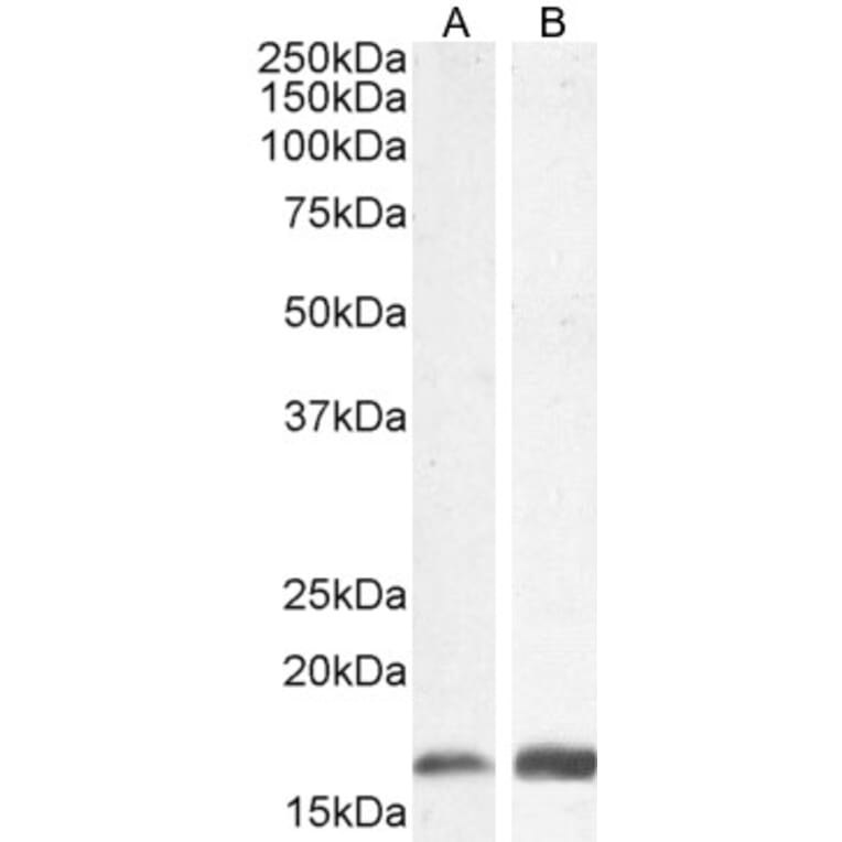
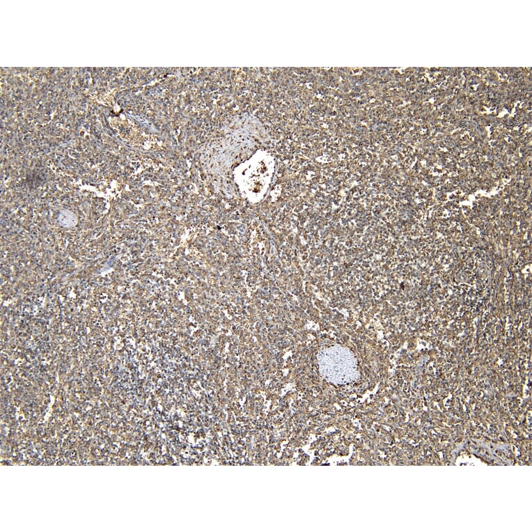
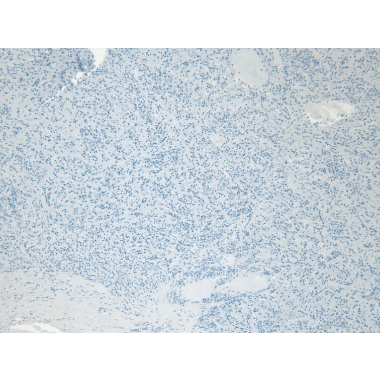
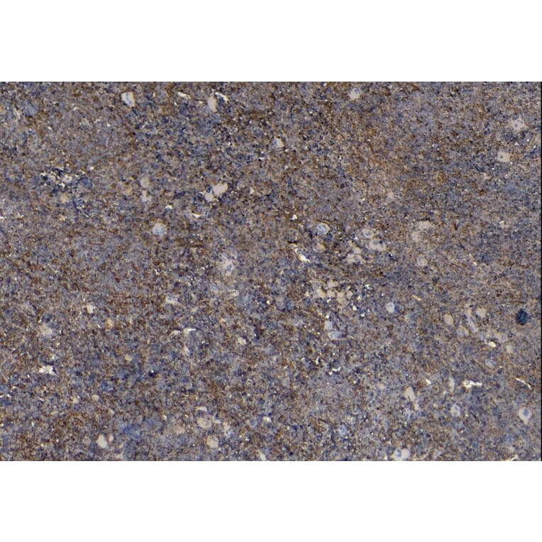
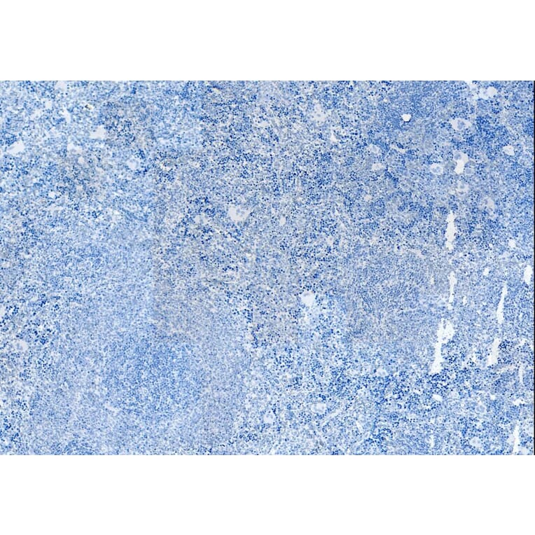







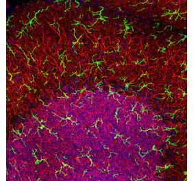
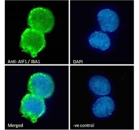
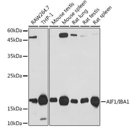
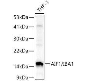
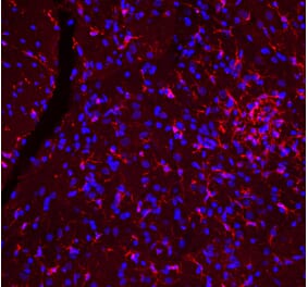
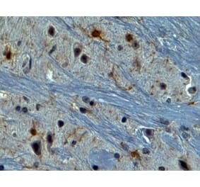
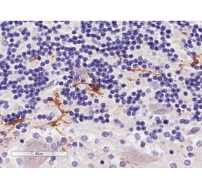
![Immunohistochemistry - Anti-Iba1 Antibody [AIF1/1909] - BSA and Azide free (A248417) - Antibodies.com](https://cdn.antibodies.com/image/catalog/251/A251599_1.jpg?profile=product_alternative)
![Immunohistochemistry - Anti-Iba1 Antibody [AIF1/1909] (A254120) - Antibodies.com](https://cdn.antibodies.com/image/catalog/248/A248417_1.jpg?profile=product_alternative)
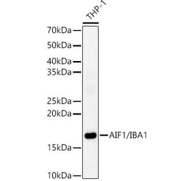
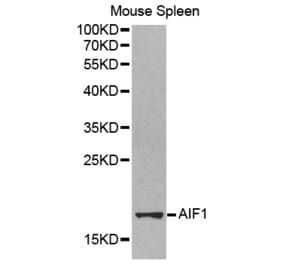
![Immunohistochemistry - Anti-Iba1 Antibody [rAIF1/1909] - BSA and Azide free (A248418) - Antibodies.com](https://cdn.antibodies.com/image/catalog/251/A251600_1.jpg?profile=product_alternative)
![Immunohistochemistry - Anti-Iba1 Antibody [AIF1/2493] - BSA and Azide free (A248419) - Antibodies.com](https://cdn.antibodies.com/image/catalog/251/A251601_1.jpg?profile=product_alternative)