

Unconjugated
Episodic ataxia type 2 (EA2) is an inherited neurological disorder, where patients suffer from chronic ataxia and severe episodes of motor dysfunction exhibited as dystonia. Despite other factors, physical and emotional stress triggers those episodes reliably in both human and mice. We used the well-established EA2 mouse model tottering to explore the cerebellar adrenergic receptor (AR) involvement in stress-induced dystonic attacks. We found that a1-ARs in cerebellar Purkinje cells (PCs) are activated by norepinephrine (NE), differentially expressed and required for initiation of dystonia, while a2-ARs are not. Moreover, pharmacological blockade and shRNA-induced knock down of cerebellar a1D-ARs was sufficient to effectively prevent stress-induced dystonia in homozygous totteringtg/tg mice but had no impact on ataxia amelioration. In vivo recordings and live calcium (Ca2+) imaging of PCs demonstrated that a1D-AR blockade successfully protects PCs from NE-mediated erratic firing patterns through decreased release of Ca2+ from intracellular stores, thus preventing stress-induced dystonia. Together, our data show the modulatory effects of NE on dystonia severity and suggest a predominant role of cerebellar a1D-ARs in the formation of stress-induced dystonia in totteringtg/tg mice.
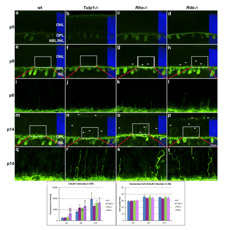
Mutations in tubby like protein 1 gene (TULP1) are causative of early-onset recessive inherited retinal degenerations (IRDs); similarly, the Tulp1-/- mouse is also characterized by a rapid IRD. Tulp1 mRNA and protein expression was analyzed in wild type mouse retinas and expression data sets (NCBI) during early postnatal development. Comparative histology was undertaken in Tulp1-/-, rhodopsin-/- (Rho-/-) and retinal degeneration slow-/- (Rds-/-) mouse retinas. Bioinformatic analysis of predicted TULP1 interactors and IRD genes was performed. Peak expression of Tulp1 in healthy mouse retinas was detected at p8; of note, TULP1 was detected in both the outer and inner retina. Bioinformatic analysis indicated Tulp1 expression in retinal progenitor, photoreceptor and non-photoreceptor cells. While common features of photoreceptor degeneration were detected in Tulp1-/-, Rho-/-, and Rds-/- retinas, other alterations in bipolar, amacrine and ganglion cells were specific to Tulp1-/- mice. Additionally, predicted TULP1 interactors differed in various retinal cell types and new functions for TULP1 were suggested. A pilot bioinformatic analysis indicated that in a similar fashion to Tulp1, many other IRD genes were expressed in both inner and outer retinal cells at p4-p7. Our data indicate that expression of Tulp1 extends to multiple retinal cell types; lack of TULP1 may lead to primary degeneration not only of photoreceptor but also non-photoreceptor cells. Predicted interactors suggest widespread retinal functions for TULP1. Early and widespread expression of TULP1 and some other IRD genes in both the inner and outer retina highlights potential hurdles in the development of treatments for these IRDs.
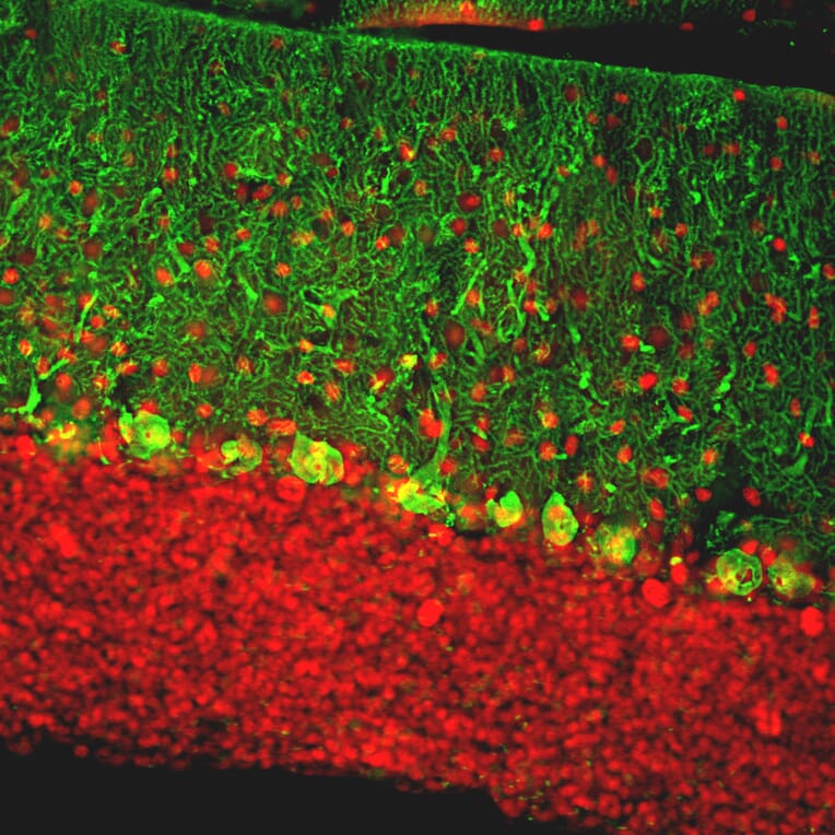
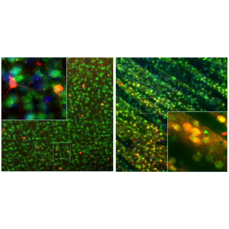
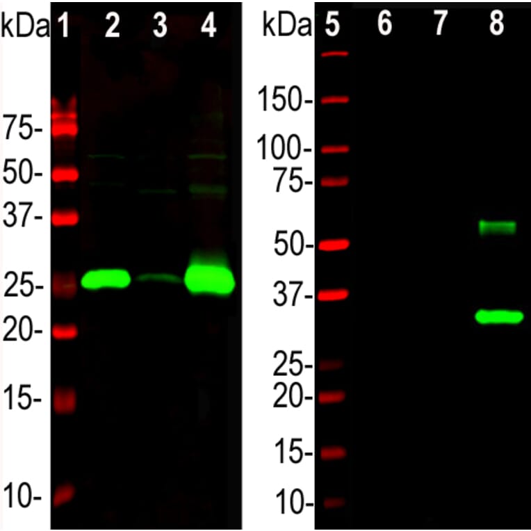
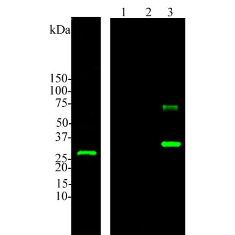
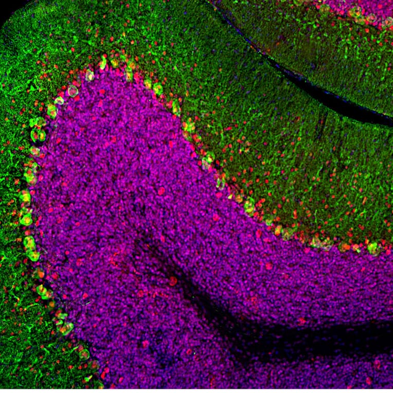
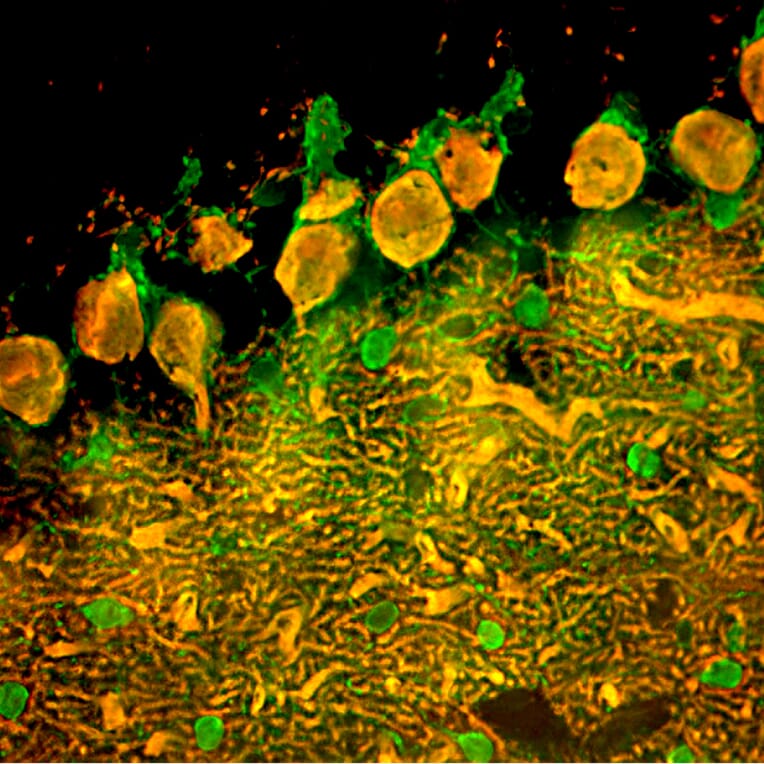
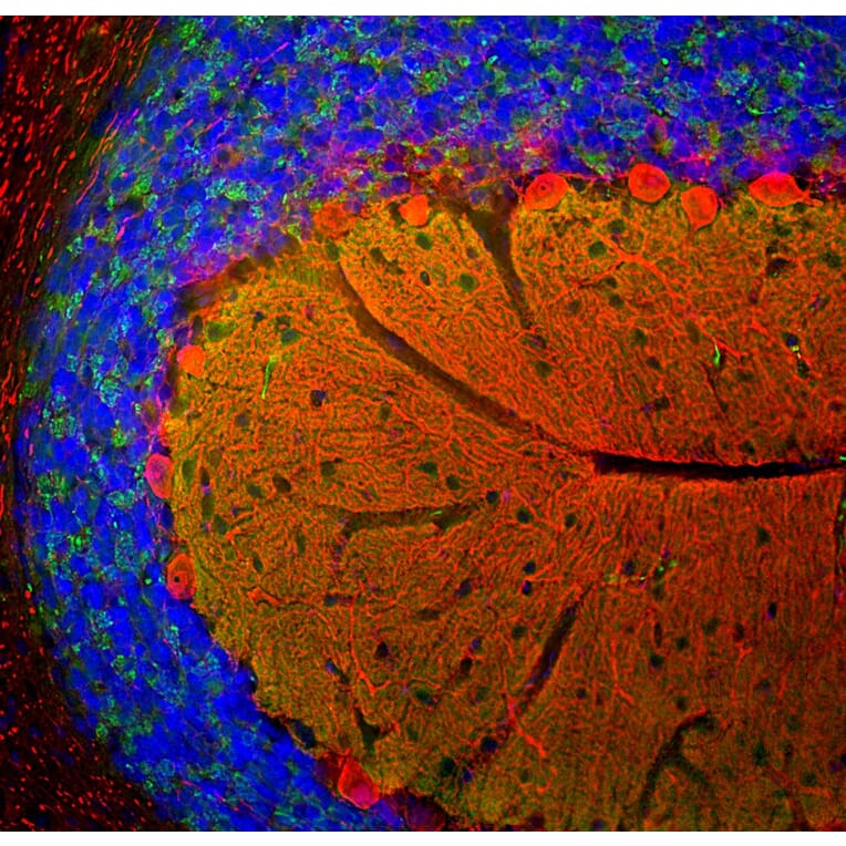
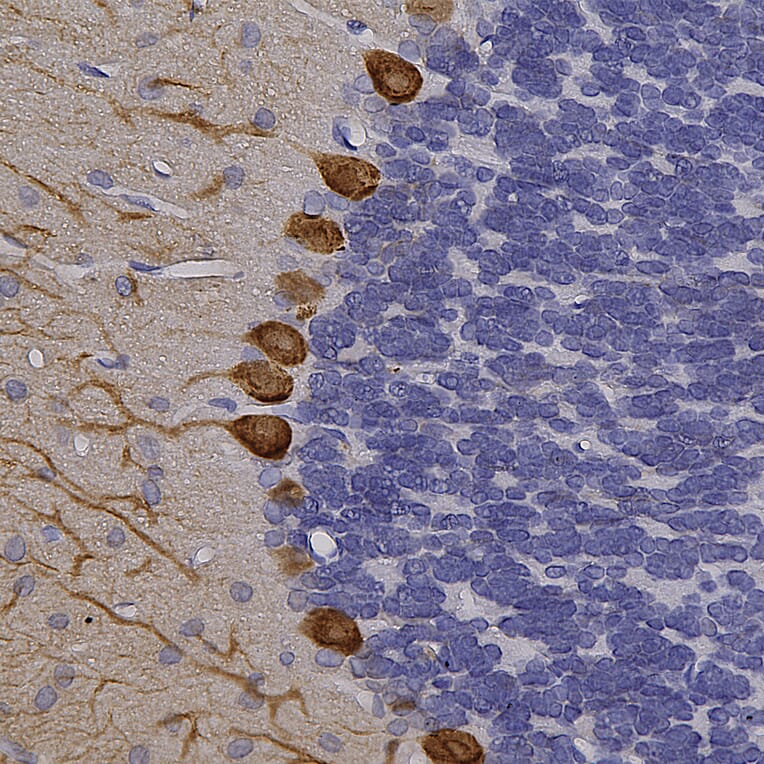
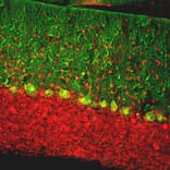
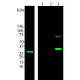
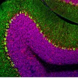
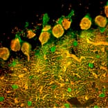
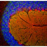
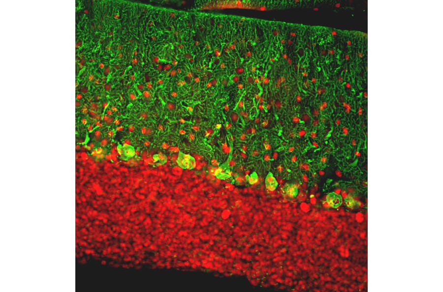
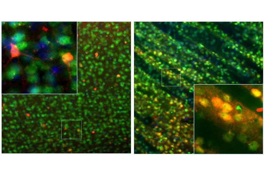
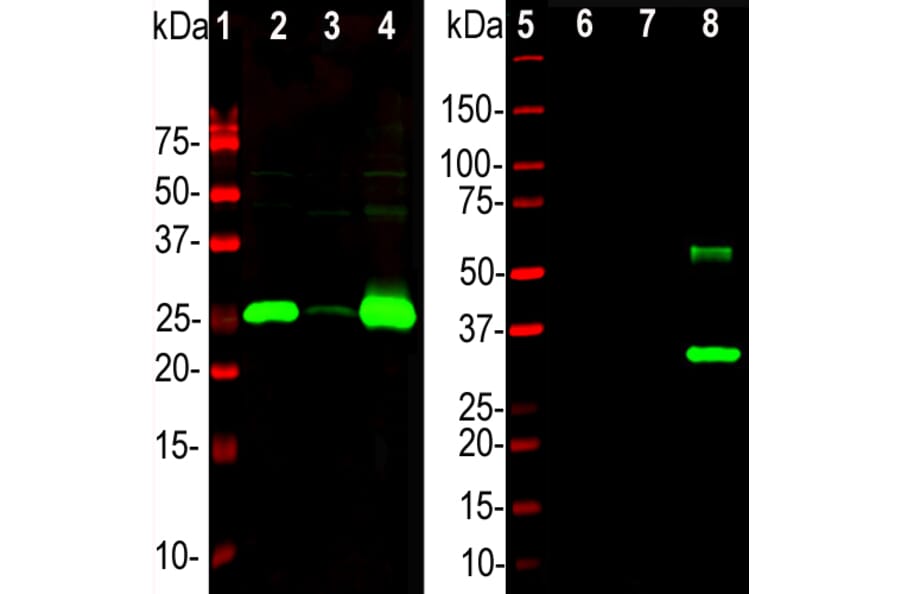
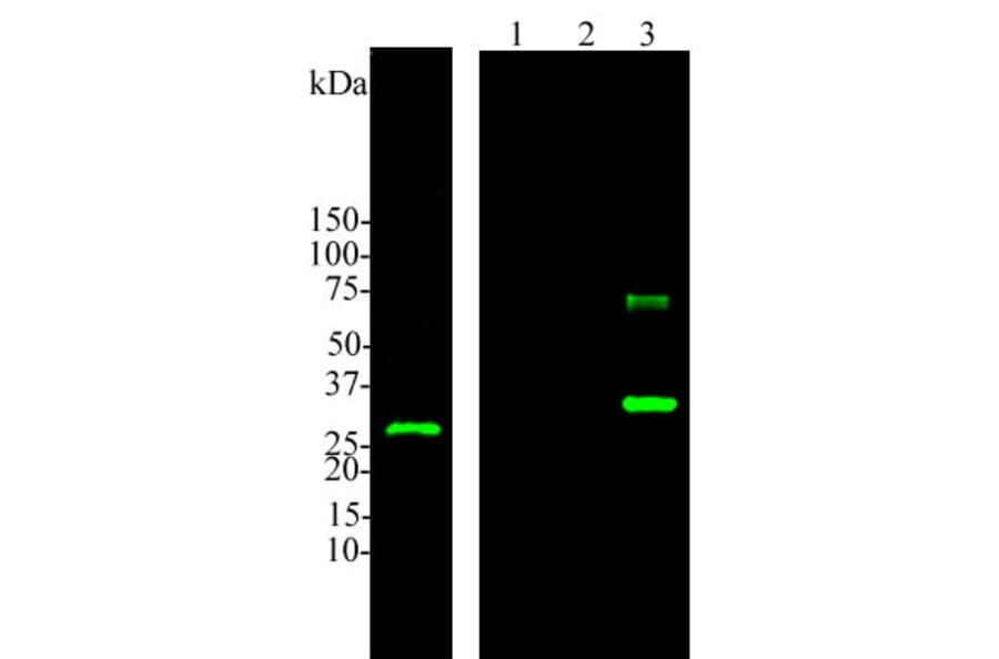
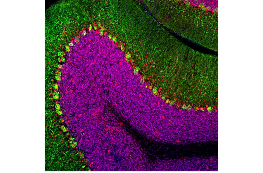
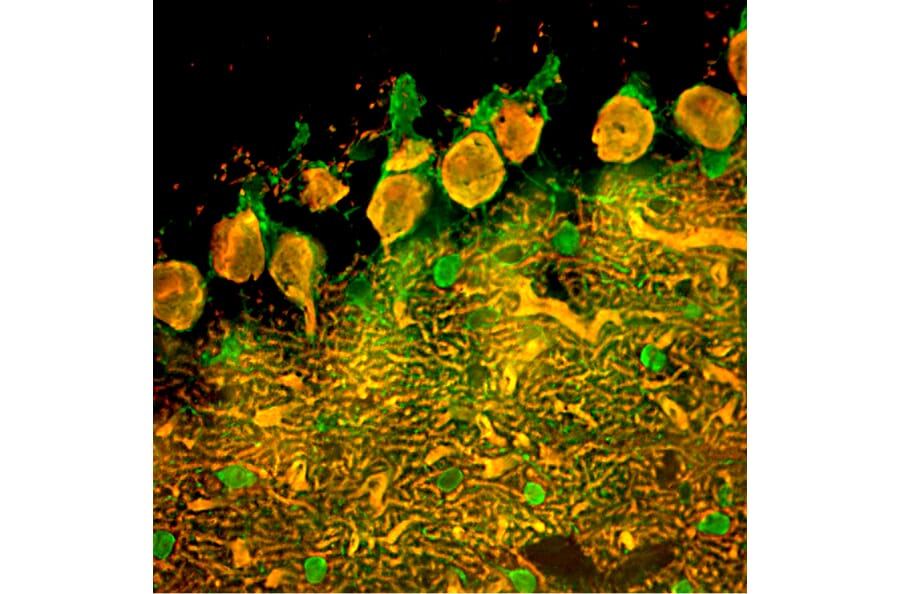
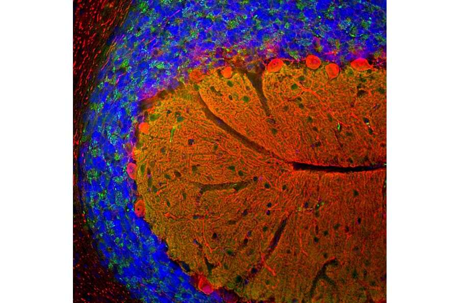
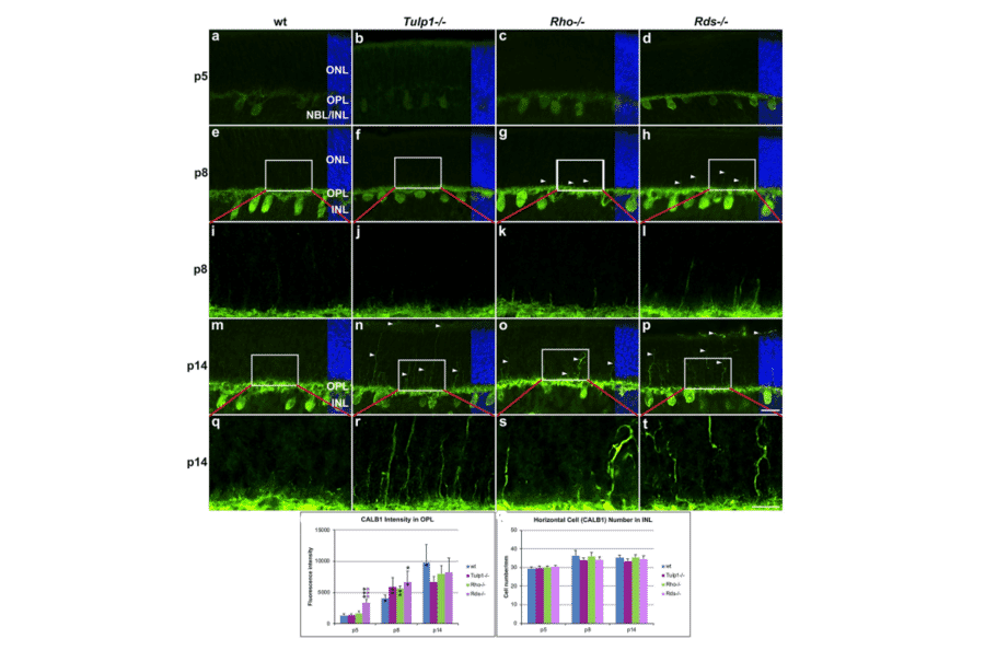

![Immunofluorescence - Anti-Calbindin Antibody [4H7] (A85360) - Antibodies.com](https://cdn.antibodies.com/image/catalog/85/A85360_1.jpg?profile=product_alternative)
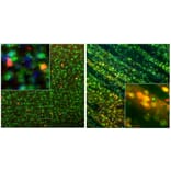
![Immunofluorescence - Anti-Calbindin Antibody [5A9] (A85362) - Antibodies.com](https://cdn.antibodies.com/image/catalog/85/A85362_1.jpg?profile=product_alternative)
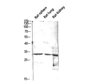
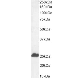
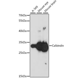
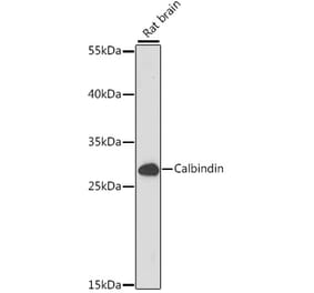
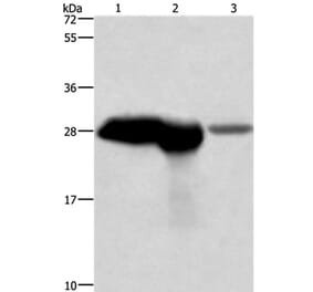
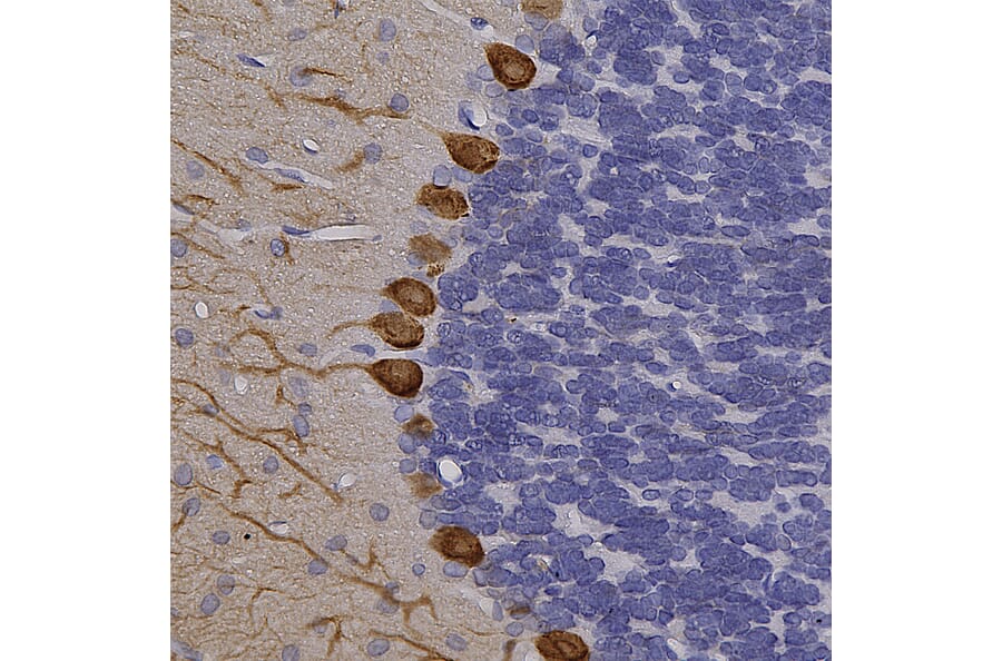
![Immunohistochemistry - Anti-Calbindin Antibody [CALB1/2782] - BSA and Azide free (A250370) - Antibodies.com](https://cdn.antibodies.com/image/catalog/253/A253550_1.jpg?profile=product_alternative)
![Immunohistochemistry - Anti-Calbindin Antibody [CALB1/3333] - BSA and Azide free (A253551) - Antibodies.com](https://cdn.antibodies.com/image/catalog/253/A253551_1.jpg?profile=product_alternative)
![Immunohistochemistry - Anti-Calbindin Antibody [CALB1/3333] (A250371) - Antibodies.com](https://cdn.antibodies.com/image/catalog/250/A250371_1.jpg?profile=product_alternative)
![Immunohistochemistry - Anti-Calbindin Antibody [CALB1/2782] (A251213) - Antibodies.com](https://cdn.antibodies.com/image/catalog/250/A250370_1.jpg?profile=product_alternative)
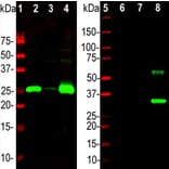
![SDS-PAGE - Anti-Calbindin Antibody [CALB1/2364] (A251249) - Antibodies.com](https://cdn.antibodies.com/image/catalog/250/A250369_1.jpg?profile=product_alternative)
![SDS-PAGE - Anti-Calbindin Antibody [CALB1/2364] - BSA and Azide free (A250369) - Antibodies.com](https://cdn.antibodies.com/image/catalog/253/A253549_1.jpg?profile=product_alternative)