Primary Antibodies
Secondary Antibodies
Proteins & Peptides
ELISA Kits
About Us
Contact Us
Sign In/Register
0
ISO 9001:2015 Certified
Live Customer Support
4.5/5 on Trustpilot
100% Quality Guarantee
Home
Primary Antibodies
GFP Antibodies
Anti-GFP Antibody (A121591)
Anti-GFP Antibody (A121591)
Overview
Specifications
Images
Enlarge Image
Enlarge Image
$590
Product Datasheet
Goat polyclonal antibody to GFP for WB, IF, IHC-P and IHC-Fr.
100% Guarantee
Price Match Guarantee
Product Size:
600µg
1.5mg
Quantity:
1
2
3
4
5
6
7
8
9
10
Add To Cart
Request a Quotation
Custom or Bulk Request
Shipping Information
Freight/Packing Charges:
$40
Dispatched from St. Louis, MO.
Lead Time: 5-8 business days.
Specifications
Name
Anti-GFP Antibody
Description
Goat polyclonal antibody to GFP.
Applications
WB
,
IF
,
IHC-P
,
IHC-Fr
Dilutions
WB: 1:500-1:5,000, IF: 1:50-1:1,000, IHC-P: 1:50-1:1,000, IHC-Fr: 1:50-1:1,000
Cross Reactivity
This antibody does not cross-react with mCherry.
Immunogen
Recombinant green fluorescent protein, expressed in and purified from E. coli.
Host
Goat
Clonality
Polyclonal
Isotype
IgG
Conjugate
Unconjugated
Purification
Affinity purification.
Concentration
3 mg/ml
Product Form
Liquid
Formulation
Supplied in Phosphate Buffered Saline with 20% Glycerol and 0.05% Sodium Azide.
Storage
Shipped at 4°C. Upon delivery aliquot and store at -20°C. Avoid freeze/thaw cycles.
Isotype Controls
Goat IgG (A121671)
Suitable Secondaries
Donkey Anti-Goat IgG H&L Antibody (AP) (A300679)
Donkey Anti-Goat IgG H&L Antibody (Biotin) (A300716)
Donkey Anti-Goat IgG H&L Antibody (FITC) (A300685)
Donkey Anti-Goat IgG H&L Antibody (HRP) (A300730)
See all Anti-Goat IgG Secondaries →
Disclaimer
This product is for research use only. It is not intended for diagnostic or therapeutic use.
Scientific Validation Data
Validation Data
(2)
Enlarge Image
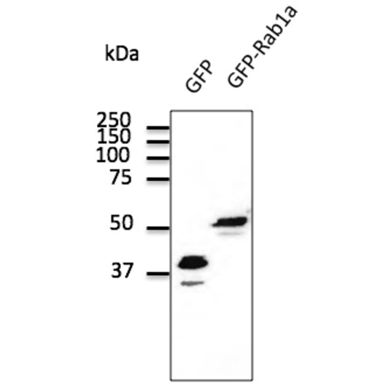
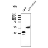
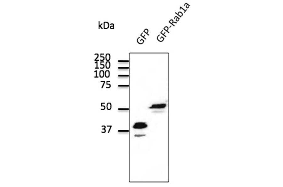
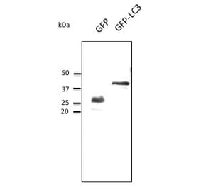
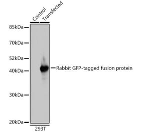
Western Blot - Anti-GFP Antibody (A121591)
Transfected 293HEK cell lysates detected with Anti-GFP Antibody at a 1:2,000 dilution. Lysates at 100µg per lane and rabbit anti-goat IgG antibody (HRP) at a 1:10,000 dilution.
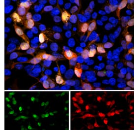
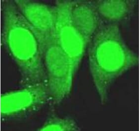
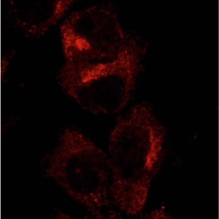
Enlarge Image
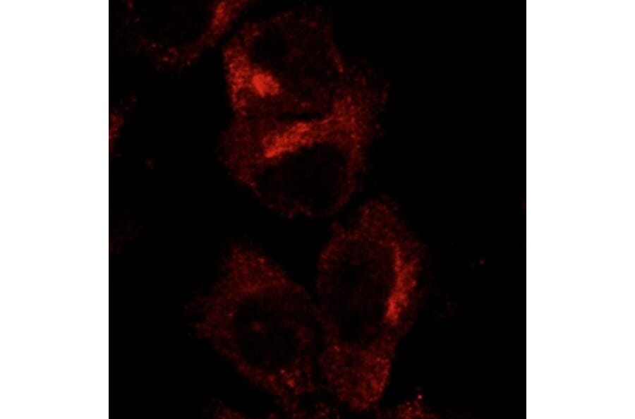
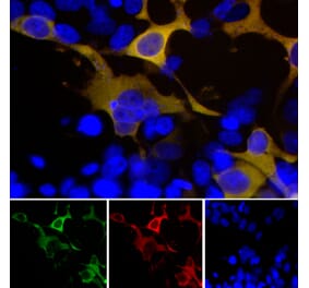
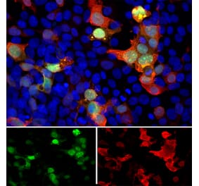
Anti-GFP Antibody (A121591)
Hepa 1-6 cells, transfected with GFP-RAB1A, stained with Anti-GFP Antibody.
Publishing research using Anti-GFP Antibody (A121591)? Please
let us know
so that we can list the citation on this page.
Most popular Anti-GFP Antibodies
(7)
A290
Anti-GFP Antibody
Rabbit polyclonal antibody to GFP for WB, ELISA, ICC/IF, Flow Cytometry, IHC-Fr, IHC-P, IP and Electron Microscopy.
(7)
A290
Anti-GFP Antibody
Rabbit polyclonal antibody to GFP for WB, ELISA, ICC/IF, Flow Cytometry, IHC-Fr, IHC-P, IP and Electron Microscopy.
Alternative products to Anti-GFP Antibody (A121591)
(3)
A86774
1 Citation
Anti-GFP Antibody
Rabbit polyclonal antibody to GFP for IP, WB and ICC.
(2)
A93400
Anti-GFP Antibody [F56-6A1.2.3]
Mouse monoclonal (F56-6A123) antibody to GFP for ELISA, WB, IP, ChIP and IHC.
(5)
A121560
1 Citation
Anti-GFP Antibody
Goat polyclonal antibody to GFP for WB, IF, IHC-P and IHC-Fr.
(5)
A85300
Anti-GFP Antibody
Chicken polyclonal antibody to GFP for WB, ICC/IF and IHC.
A85283
Anti-GFP Tag Antibody [GF28R]
Mouse monoclonal (GF28R) antibody to GFP Tag for Dot, ELISA, IP, IS and WB.
(4)
A104347
Anti-GFP Antibody
Goat polyclonal antibody to GFP for WB, ICC/IF and IHC.
(3)
A17321
Anti-GFP Antibody
Rabbit polyclonal antibody to GFP for WB and ICC/IF.
(4)
A252
1 Citation
Anti-GFP Antibody
Rabbit polyclonal antibody to GFP for WB, IP, IF and IHC.
A50024
2 Citations
Anti-GFP (Plant Specific) Antibody
Mouse monoclonal antibody to GFP (Plant Specific) for WB.
(3)
A251
Anti-GFP Antibody
Rat monoclonal (1A5) antibody to GFP for WB, IP, ICC, ChIP and ELISA.
(5)
A85298
Anti-GFP Antibody
Rabbit polyclonal antibody to GFP for WB and ICC/IF.
A93401
Anti-GFP Antibody [7A1]
Mouse monoclonal (7A1) antibody to GFP for ELISA, WB, IP, ChIP and IHC.
(3)
A86774
1 Citation
Anti-GFP Antibody
Rabbit polyclonal antibody to GFP for IP, WB and ICC.
(2)
A93400
Anti-GFP Antibody [F56-6A1.2.3]
Mouse monoclonal (F56-6A123) antibody to GFP for ELISA, WB, IP, ChIP and IHC.
(5)
A121560
1 Citation
Anti-GFP Antibody
Goat polyclonal antibody to GFP for WB, IF, IHC-P and IHC-Fr.
(5)
A85300
Anti-GFP Antibody
Chicken polyclonal antibody to GFP for WB, ICC/IF and IHC.
A85283
Anti-GFP Tag Antibody [GF28R]
Mouse monoclonal (GF28R) antibody to GFP Tag for Dot, ELISA, IP, IS and WB.
(4)
A104347
Anti-GFP Antibody
Goat polyclonal antibody to GFP for WB, ICC/IF and IHC.
(3)
A17321
Anti-GFP Antibody
Rabbit polyclonal antibody to GFP for WB and ICC/IF.
(4)
A252
1 Citation
Anti-GFP Antibody
Rabbit polyclonal antibody to GFP for WB, IP, IF and IHC.
A50024
2 Citations
Anti-GFP (Plant Specific) Antibody
Mouse monoclonal antibody to GFP (Plant Specific) for WB.
(3)
A251
Anti-GFP Antibody
Rat monoclonal (1A5) antibody to GFP for WB, IP, ICC, ChIP and ELISA.
(5)
A85298
Anti-GFP Antibody
Rabbit polyclonal antibody to GFP for WB and ICC/IF.
A93401
Anti-GFP Antibody [7A1]
Mouse monoclonal (7A1) antibody to GFP for ELISA, WB, IP, ChIP and IHC.
See all GFP Antibodies
Top


![Fluorescent image of COS1 cells due to GFP of GST-ZIPK fusion protein expressed in HEK293T cells (Right) and the same cells were immunostained using Anti-GFP Antibody [1A5], followed by Anti-Rat IgG (Texas Red) (Left). Note that fluorescence by the immunofluorescent staining using Anti-GFP Antibody [1A5] is much stronger than fluorescence due to GFP.](https://cdn.antibodies.com/image/catalog/0/A251_1.png?profile=product_alternative)