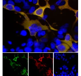
Unconjugated
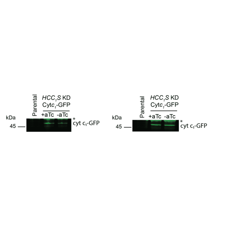
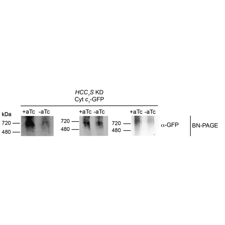
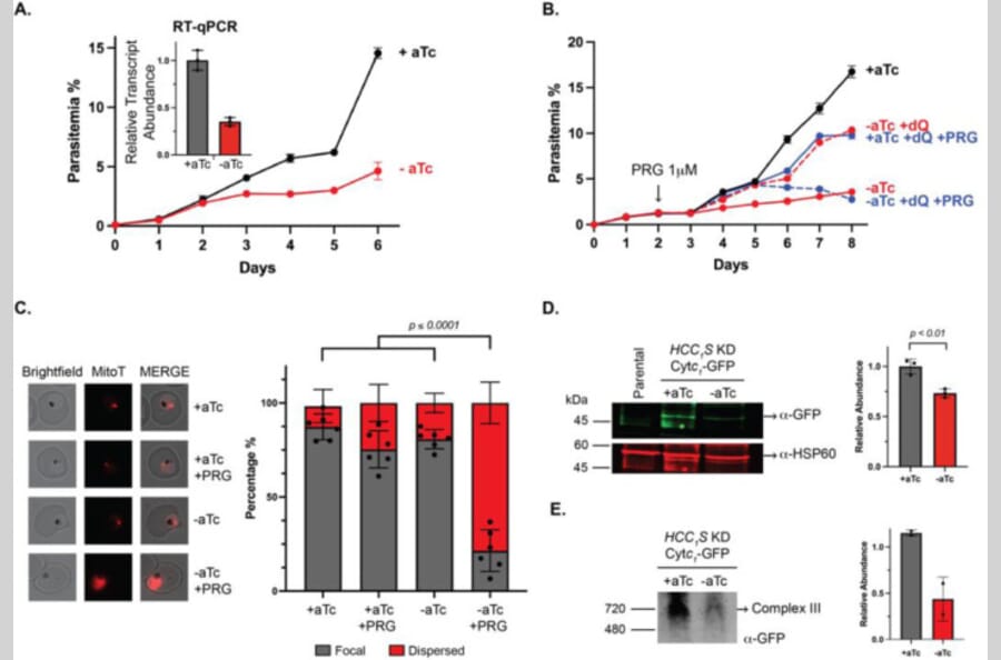
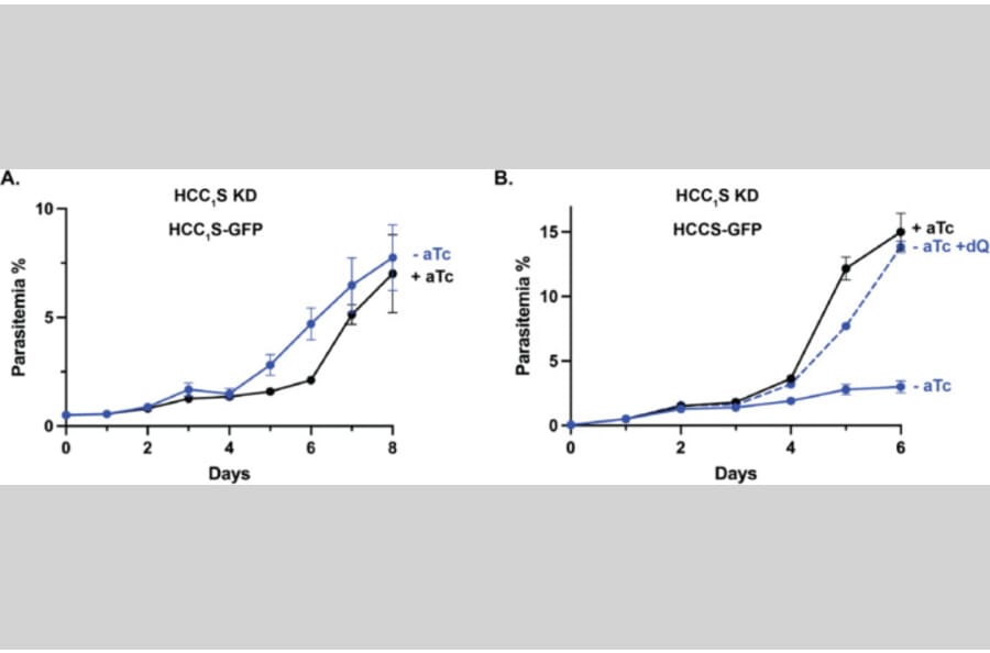
Plasmodium malaria parasites retain an essential mitochondrional electron transport chain (ETC) that is critical for growth within humans and mosquitoes and a key antimalarial drug target. ETC function requires cytochromes c and c1 that are unusual among heme proteins due to their covalent binding to heme via conserved CXXCH sequence motifs. Heme attachment to these proteins in most eukaryotes requires the mitochondrial enzyme holocytochrome c synthase (HCCS) that binds heme and the apo cytochrome to facilitate biogenesis of the mature cytochrome c or c1. Although humans encode a single bifunctional HCCS that attaches heme to both proteins, Plasmodium parasites are like yeast and encode two separate HCCS homologs thought to be specific for heme attachment to cyt c (HCCS) or cyt c1 (HCC1S). To test the function and specificity of P. falciparum HCCS and HCC1S, we used CRISPR/Cas9 to tag both genes for conditional expression. HCC1S knockdown selectively impaired cyt c1 biogenesis and caused lethal ETC dysfunction that was not reversed by over-expression of HCCS. Knockdown of HCCS caused a more modest growth defect but strongly sensitized parasites to mitochondrial depolarization by proguanil, revealing key defects in ETC function. These results and prior heterologous studies in E. coli of cyt c hemylation by P. falciparum HCCS and HCC1S strongly suggest that both homologs are essential for mitochondrial ETC function and have distinct specificities for biogenesis of cyt c and c1, respectively, in parasites. This study lays a foundation to develop novel strategies to selectively block ETC function in malaria parasites.
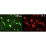


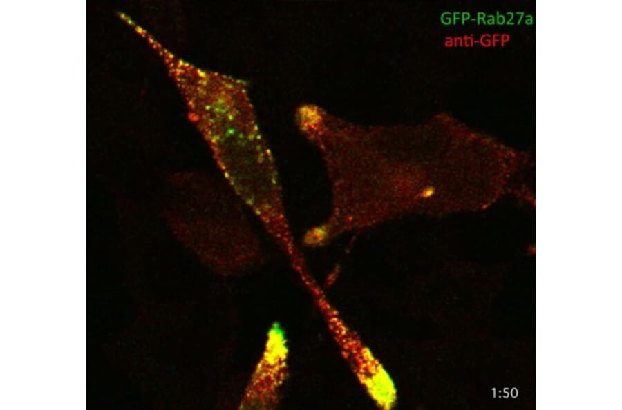

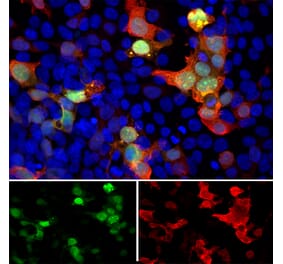
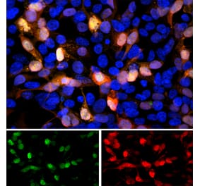
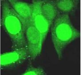
![Fluorescent image of COS1 cells due to GFP of GST-ZIPK fusion protein expressed in HEK293T cells (Right) and the same cells were immunostained using Anti-GFP Antibody [1A5], followed by Anti-Rat IgG (Texas Red) (Left). Note that fluorescence by the immunofluorescent staining using Anti-GFP Antibody [1A5] is much stronger than fluorescence due to GFP.](https://cdn.antibodies.com/image/catalog/0/A251_1.png?profile=product_alternative)