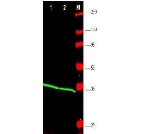
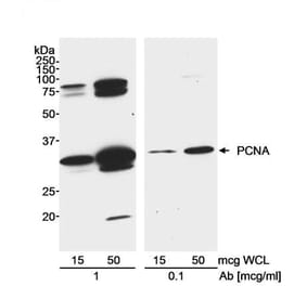
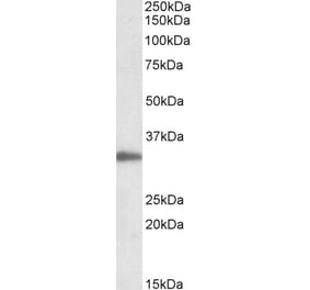
This antibody detects endogenous levels of PCNA and does not cross-react with related proteins
Applications
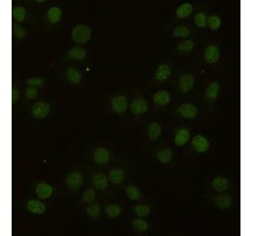
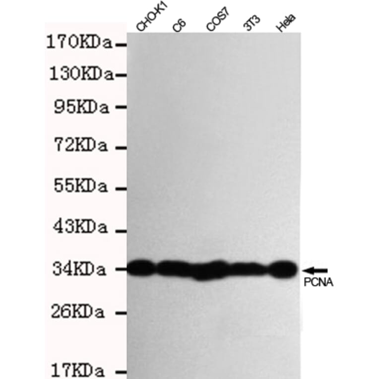
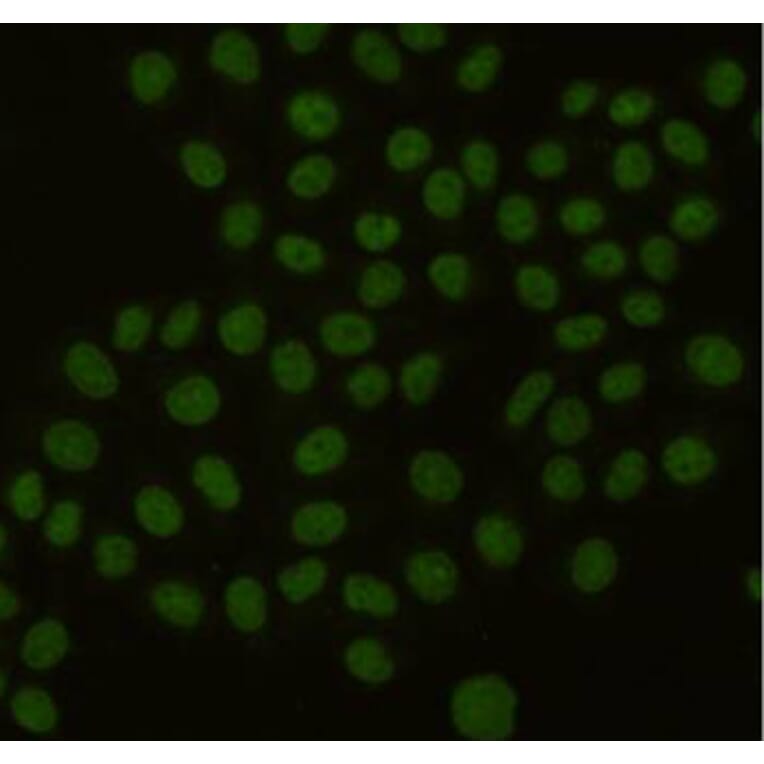
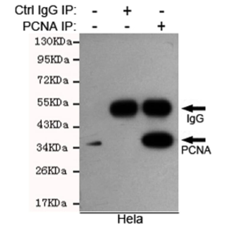
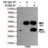
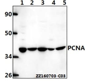
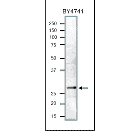
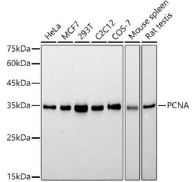
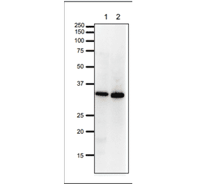
WB, IP, ICC
Reactivity
Human, Mouse, Rat, Monkey, Hamster
Immunogen
Recombinant full length Human PCNA.
Host
Mouse
Clonality
Monoclonal
Conjugate
Unconjugated
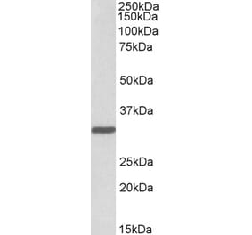
Molecular Weight
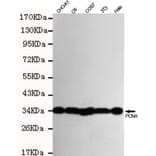
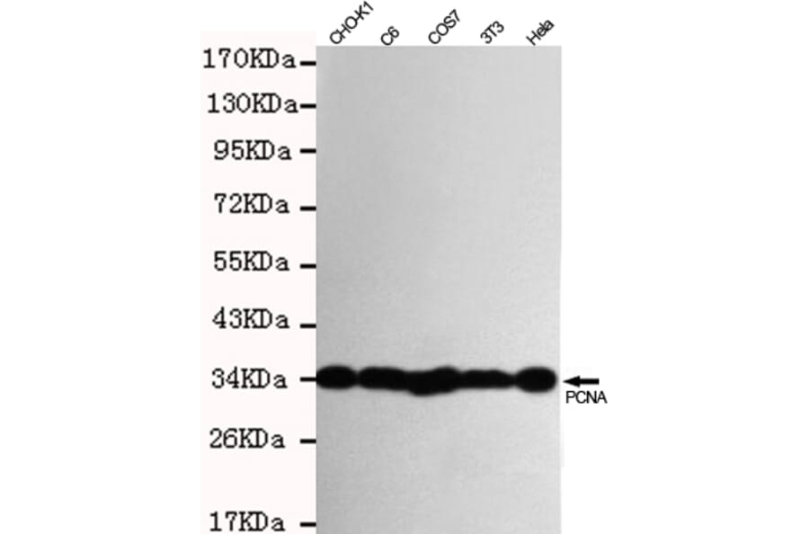
Predicted band size:36KDaObserved band size:36KDa
Purity
The antibody was affinity-purified from mouse ascites by affinity-chromatography using epitope-specific immunogen and the purity is > 95% (by SDS-PAGE).
Product Form
Purified mouse monoclonal in buffer containing 0.1M Tris-Glycine (pH 7.4, 150 mM NaCl) with 0.2% sodium azide, 50%,glycerol


![Flow Cytometry - Anti-PCNA Antibody [PC10] (A86878)](https://cdn.antibodies.com/image/catalog/86/A86878_1.jpg?profile=product_alternative)
![SDS-PAGE - Anti-PCNA Antibody [PC10] (A281549) - Antibodies.com](https://cdn.antibodies.com/image/catalog/281/A281549_3.jpg?profile=product_alternative)
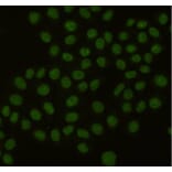
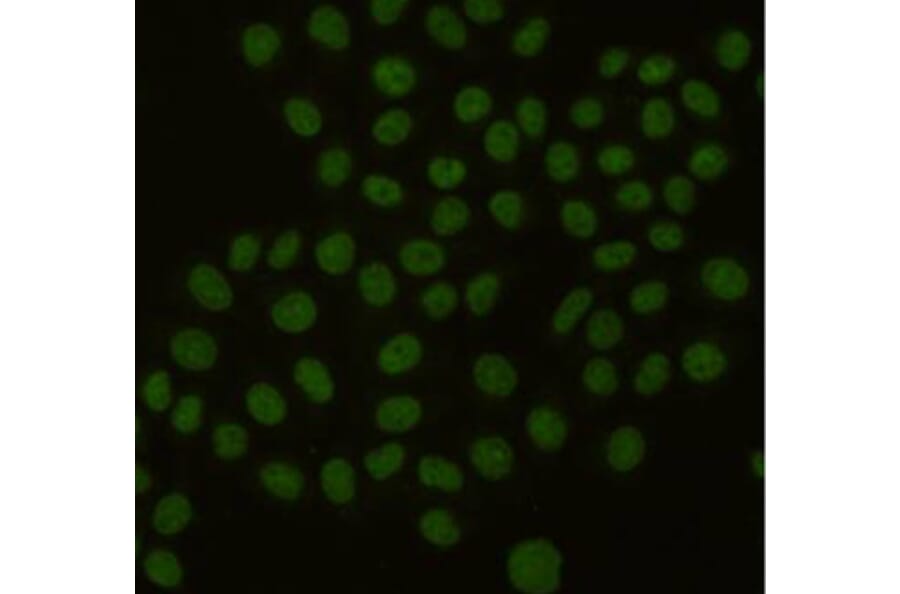
![Immunohistochemistry - Anti-PCNA Antibody [PC10] (A252744) - Antibodies.com](https://cdn.antibodies.com/image/catalog/249/A249578_1.jpg?profile=product_alternative)
![Immunohistochemistry - Anti-PCNA Antibody [PC10] - BSA and Azide free (A249578) - Antibodies.com](https://cdn.antibodies.com/image/catalog/252/A252758_1.jpg?profile=product_alternative)
![Immunohistochemistry - Anti-PCNA Antibody [SPM350] - BSA and Azide free (A249579) - Antibodies.com](https://cdn.antibodies.com/image/catalog/252/A252759_1.jpg?profile=product_alternative)
![Immunohistochemistry - Anti-PCNA Antibody [SPM350] (A252760) - Antibodies.com](https://cdn.antibodies.com/image/catalog/249/A249579_1.jpg?profile=product_alternative)