
Unconjugated
Background: This study examines the impact of a light-cycle shift regimen on corneal and conjunctival tissues in menopausal rats and evaluates the protective role of combined hormone therapy.
Methods: Twenty-four menopausal female albino rats were randomly assigned to three groups (n = 8) following a 10-day acclimatization period. Group 1 (Control+Saline) was maintained under a 12:12 light/dark cycle. Light-cycle shift regimen was induced in Groups 2 and 3 using a rotating 7-day light-exposure sequence repeated over 21 days; this protocol consisted of 24 h of continuous light, 72 h of inverted dark-light timing, and 72 h of standard light-dark conditions. Groups 1 and 2 received saline, while Group 3 received 17ß-Estradiol and drospirenone daily via oral gavage. After 31 days, eyes were enucleated for histological and immunohistochemical analyses of corneal, conjunctival, and palpebral tissues, including caspase-3 (Cas-3), tumor necrosis factor-alpha (TNF-a), and PERIOD-2 (PER2) expression.
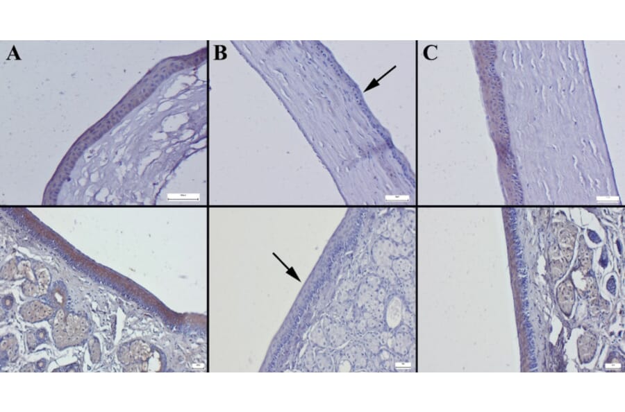
Results: Light-cycle shift regimen (Group 2) significantly increased corneal thickness (p < 0.001), conjunctival inflammation, and vascular congestion, with marked upregulation of Cas-3 and TNF-a and downregulation of PER2. Hormone therapy (Group 3) attenuated these effects, showing reduced corneal edema, diminished inflammatory infiltration, and partial normalization of molecular markers.
Conclusions: Shifting light-dark cycles may aggravate inflammatory and apoptotic changes in the ocular surface during menopause. Estrogen-progestin therapy attenuates these alterations by modulating the expression of the circadian-associated protein PER2 and maintaining structural integrity. These findings suggest that hormone therapy may offer potential benefits for preserving ocular surface homeostasis in menopausal women experiencing sleep or circadian rhythm disturbances.
Circadian rhythms are biological processes that occur in approximately 24-hour cycles in organisms. Disruption of circadian rhythms (CD) is thought to have adverse effects on many organs, including the eyes. The aim of this study was to evaluate the effects of circadian disruption on the cornea and conjunctiva, as well as the impact of two forms of omega-3 supplementation on the ocular surface, using a rat model of circadian disruption. This experimental study included 32 female Wistar albino rats, which were divided into four groups. The control group (Group 1) was maintained under normal feeding and sleeping conditions without any disruption. Circadian disruption was induced in the other three groups, which were administered saline (Group 2), fish oil (Group 3), or flaxseed oil (Group 4) via oral gavage. On the 31st day, all rats were euthanized, and corneal, conjunctival, and palpebral tissues were collected from both eyes through enucleation. Histological examination and immunohistochemical analysis of Caspase-3 (Cas-3), tumor necrosis factor-alpha (TNF-a), and PERIOD-2 (PER-2) were performed on the corneal and conjunctival tissues. Group 2 exhibited significantly thicker corneas compared to Group 1 (P < 0.001). Additionally, hyperemia, inflammatory cell infiltration in the conjunctiva, higher expression levels of Cas-3 and TNF-a, and decreased PER-2 expression were observed in the corneal and conjunctival tissues of Group 2. These pathological changes were minimal or absent in Groups 3 and 4. Notably, Group 3 showed better amelioration of these alterations compared to Group 4. Disruption of circadian rhythms can have a negative impact on the cornea and conjunctiva. Omega-3 supplementation demonstrated a significant protective effect against ocular tissue damage induced by circadian disruption. Fish oil was more effective than flaxseed oil in reducing corneal thickening, inflammation, and apoptotic marker expression, highlighting its potential as a therapeutic intervention for circadian rhythm-related ocular pathologies.