Unconjugated
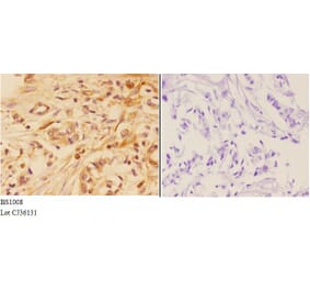
Amplified in breast cancer 1 (AIB1) is a member of p160 steroid receptor coactivator (SRC) family that mediates the transcriptional activities of nuclear receptors and other transcription factors. It acts as a major oncogene in diverse cancers, whereas biological function of AIB1 in gastric cancer remains largely unclear. This study was designed to explore the role of AIB1 in gastric tumorigenesis and its potential as a useful prognostic marker and therapeutic target in this cancer. Our data demonstrated that AIB1 was significantly up-regulated in gastric cancer tissues as compared with control subjects. Moreover, AIB1 amplification was found in 47 of 133 (35.3%) gastric cancer cases, but not in control subjects. AIB1 amplification was positively associated with its protein expression, and was significantly correlated with poor patient survival. AIB1 knockdown in gastric cancer cells dramatically inhibited cell proliferation, invasiveness and tumorigenic potential in nude mice, and induced cell cycle arrest and apoptosis. Mechanically, AIB1 promotes gastric cancer cell proliferation, survival and invasiveness through modulating major signaling pathways such as ErbB and Wnt/β-catenin pathways. Collectively, these findings suggest that AIB1 plays an important role in the pathogenesis of gastric cancer and represents a potential prognostic marker and therapeutic target for this cancer.
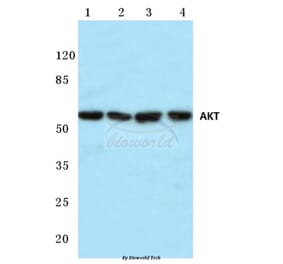
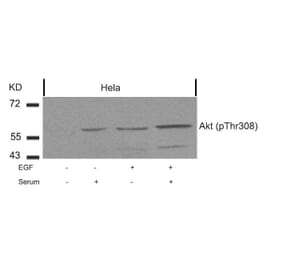
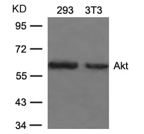
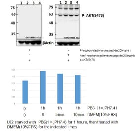
Our previous study revealed that Type II cGMP-dependent protein kinase (PKG II) inhibits epidermal growth factor (EGF)-induced MAPK/ERK and MAPK/JNK-mediated signal transduction through the inhibition of the phosphorylation/activation of the EGF receptor (EGFR). As EGFR also mediates several other signal transduction pathways besides MAPK-mediated pathways, the present study was designed to investigate whether PKG II was able to inhibit EGF/EGFR-induced phosphatidylinositol-3-kinase (PI3K)/Akt-mediated signal transduction. The AGS human gastric cancer cell line was infected with adenoviral constructs encoding a cDNA of PKG II (Ad-PKG II) to increase the expression of PKG II, and treated with 8-pCPT-cGMP to activate the enzyme. Western blotting was used to detect the phosphorylation/activation of the key components of the signal transduction pathway, including EGFR, PI3K, Akt, mTOR and NF-κB. The levels of apoptosis-related proteins, including Bax, Bcl-2, caspase 9 and DNA fragment factor (DFF), were also determined by western blotting. Terminal deoxynucleotidyl transferase-mediated dUTP nick end-labeling staining was used to detect the apoptosis of the AGS cells. The results revealed that EGF treatment increased the phosphorylation (activation) of EGFR, PI3K, Akt and mTOR, and increased the nuclear localization (activation) of NF-κB. EGF treatment also reduced the apoptosis of the AGS cells and increased the expression of the anti-apoptotic protein, Bcl-2, but had no effect on the expression of the pro-apoptotic protein, Bax, and did not alter the levels of caspase 9 and DFF. Increasing the PKG II activity of AGS cells by infecting them with Ad-PKG II and stimulating them with 8-pCPT-cGMP inhibited the EGF-induced activation of EGFR, PI3K, Akt, mTOR and NF-κB; caused an increase in caspase 9 breakdown (activation) and DFF levels; and reversed the anti-apoptotic effect of EGF. The results suggest that PKG II may also inhibit EGF-induced signal transduction of PI3K/Akt-mediated pathways, and further confirm that PKG II is able to block the activation of EGFR.


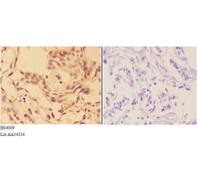
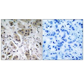
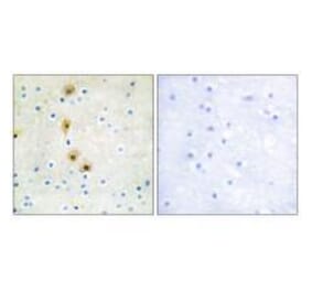
![Immunohistochemistry - Anti-AKT Antibody [RM316] (A121372) - Antibodies.com](https://cdn.antibodies.com/image/catalog/121/A121394_1.png?profile=product_alternative)
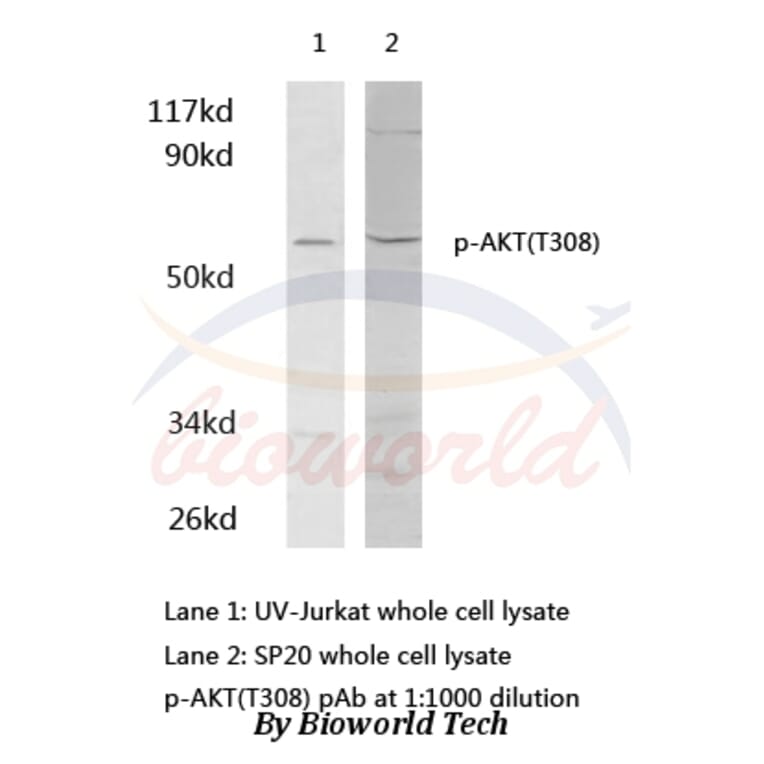
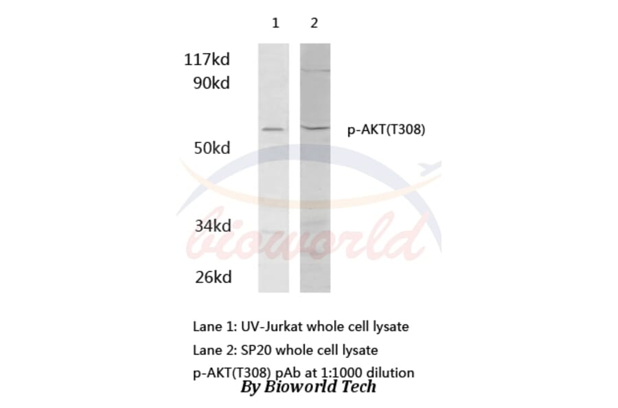
![Western Blot - Anti-AKT (phospho Ser473) Antibody [RM251] (A121248) - Antibodies.com](https://cdn.antibodies.com/image/catalog/121/A121248_1.png?profile=product_alternative)