Immunohistochemistry of a section of rat facial nucleus 7 days following axotomy using Anti-Alpha-Internexin Antibody. These neurons are capable of regenerating their axons and also, concomitant with regeneration, strongly upregulate a-internexin in their perikarya. Other central neurons which are not able to regenerate their axons do not upregulate this protein after axotomy and untreated facial neurons normally show only very low levels of a-internexin. Both findings suggest that a-internexin has a role in axonal regeneration.
Rat facial nucleus before (left) and after (right) facial nerve lesion. Note the profound upregulation of a-internexin in the facial nucleus motor neurons.
Western Blot - Anti-alpha Internexin Antibody [2E3] (A85448)
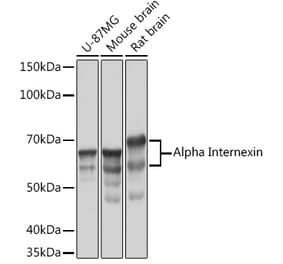
Western blot analysis of different tissue lysates using Anti-alpha Internexin Antibody [2E3] (A85448), at a dilution of 1:10,000, in red. The lanes contain samples of: [Lane 1] Protein standards, in red, [Lane 2] rat brain, [Lane 3] rat spinal cord, [Lane 4] mouse brain, [Lane 5] mouse spinal cord, and [Lane 6] cow spinal cord lysates. The Anti-alpha Internexin Antibody [2E3] (A85448) reveals the alpha Internexin protein with an apparent molecular weight of 64-66 kDa, with some variability between species.
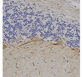
Immunohistochemistry analysis of a formalin fixed paraffin embedded human cerebellum section with Anti-alpha Internexin Antibody [2E3] (A85448) at a dilution of 1:1,000 detected in DAB (brown) using the Vector Labs ImmPRESS method and reagents with citra buffer retrieval. Counterstained with Hematoxylin (blue). The 2E3 a-Internexin antibody strongly labels neuronal processes in both rat and human brain. Note: this antibody performs well in testing with both 4% PFA and standard NBF fixed tissues.
Western Blot - Anti-alpha Internexin Antibody [2E3] (A85448)
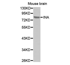
Western blot of Anti-Alpha-Internexin Antibody on recombinant full length human a-internexin at a range of concentrations from 10ng, 25ng and 50ng per gel lane as indicated. Note the strong signal even in the lane containing only 10ng of protein. The “St” lane contains SDS-PAGE molecular weight standards of the indicated size.
Western Blot - Anti-alpha Internexin Antibody [2E3] (A85448)
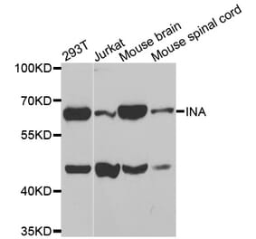
Western blot analysis of different tissue lysates using Anti-Calretinin Antibody (A104312), at a dilution of 1:1,000, in green. The lanes contain: [Lane 1] protein standard (red), [Lane 2] rat brain, [Lane 3] rat spinal cord, [Lane 4] mouse brain, [Lane 5] mouse spinal cord, and [Lane 6] cow spinal cord. A band at 29kDa corresponds to Calretinin protein. The same blot was simultaneously probed with Anti-alpha Internexin Antibody [2E3] (A85448), at a dilution of 1:10,000, in red, that reveals the alpha Internexin protein with apparent molecular weight of 66kDa.


![Immunohistochemistry - Anti-alpha Internexin Antibody [2E3] (A85448) - Antibodies.com](https://cdn.antibodies.com/image/catalog/85/A85448_1.jpg?profile=product_top)
![Immunohistochemistry - Anti-alpha Internexin Antibody [2E3] (A85448) - Antibodies.com](https://cdn.antibodies.com/image/catalog/85/A85448_2.jpg?profile=product_top)
![Western Blot - Anti-alpha Internexin Antibody [2E3] (A85448) - Antibodies.com](https://cdn.antibodies.com/image/catalog/85/A85448_3.jpg?profile=product_top)
![Immunohistochemistry - Anti-alpha Internexin Antibody [2E3] (A85448) - Antibodies.com](https://cdn.antibodies.com/image/catalog/85/A85448_4.jpg?profile=product_top)
![Western Blot - Anti-alpha Internexin Antibody [2E3] (A85448) - Antibodies.com](https://cdn.antibodies.com/image/catalog/85/A85448_5.jpg?profile=product_top)
![Western Blot - Anti-alpha Internexin Antibody [2E3] (A85448) - Antibodies.com](https://cdn.antibodies.com/image/catalog/85/A85448_6.jpg?profile=product_top)
![Immunohistochemistry - Anti-alpha Internexin Antibody [2E3] (A85448) - Antibodies.com](https://cdn.antibodies.com/image/catalog/85/A85448_1.jpg?profile=product_top_thumb)
![Immunohistochemistry - Anti-alpha Internexin Antibody [2E3] (A85448) - Antibodies.com](https://cdn.antibodies.com/image/catalog/85/A85448_2.jpg?profile=product_top_thumb)
![Western Blot - Anti-alpha Internexin Antibody [2E3] (A85448) - Antibodies.com](https://cdn.antibodies.com/image/catalog/85/A85448_3.jpg?profile=product_top_thumb)
![Immunohistochemistry - Anti-alpha Internexin Antibody [2E3] (A85448) - Antibodies.com](https://cdn.antibodies.com/image/catalog/85/A85448_4.jpg?profile=product_top_thumb)
![Western Blot - Anti-alpha Internexin Antibody [2E3] (A85448) - Antibodies.com](https://cdn.antibodies.com/image/catalog/85/A85448_5.jpg?profile=product_top_thumb)
![Western Blot - Anti-alpha Internexin Antibody [2E3] (A85448) - Antibodies.com](https://cdn.antibodies.com/image/catalog/85/A85448_6.jpg?profile=product_top_thumb)
![Immunohistochemistry - Anti-alpha Internexin Antibody [2E3] (A85448) - Antibodies.com](https://cdn.antibodies.com/image/catalog/85/A85448_1.jpg?profile=product_image)
![Immunohistochemistry - Anti-alpha Internexin Antibody [2E3] (A85448) - Antibodies.com](https://cdn.antibodies.com/image/catalog/85/A85448_2.jpg?profile=product_image)
![Western Blot - Anti-alpha Internexin Antibody [2E3] (A85448) - Antibodies.com](https://cdn.antibodies.com/image/catalog/85/A85448_3.jpg?profile=product_image)
![Immunohistochemistry - Anti-alpha Internexin Antibody [2E3] (A85448) - Antibodies.com](https://cdn.antibodies.com/image/catalog/85/A85448_4.jpg?profile=product_image)
![Western Blot - Anti-alpha Internexin Antibody [2E3] (A85448) - Antibodies.com](https://cdn.antibodies.com/image/catalog/85/A85448_5.jpg?profile=product_image)
![Western Blot - Anti-alpha Internexin Antibody [2E3] (A85448) - Antibodies.com](https://cdn.antibodies.com/image/catalog/85/A85448_6.jpg?profile=product_image)
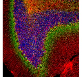
![Immunofluorescence - Anti-alpha Internexin Antibody [1D2] (A85447) - Antibodies.com](https://cdn.antibodies.com/image/catalog/85/A85447_1.jpg?profile=product_alternative)