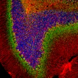
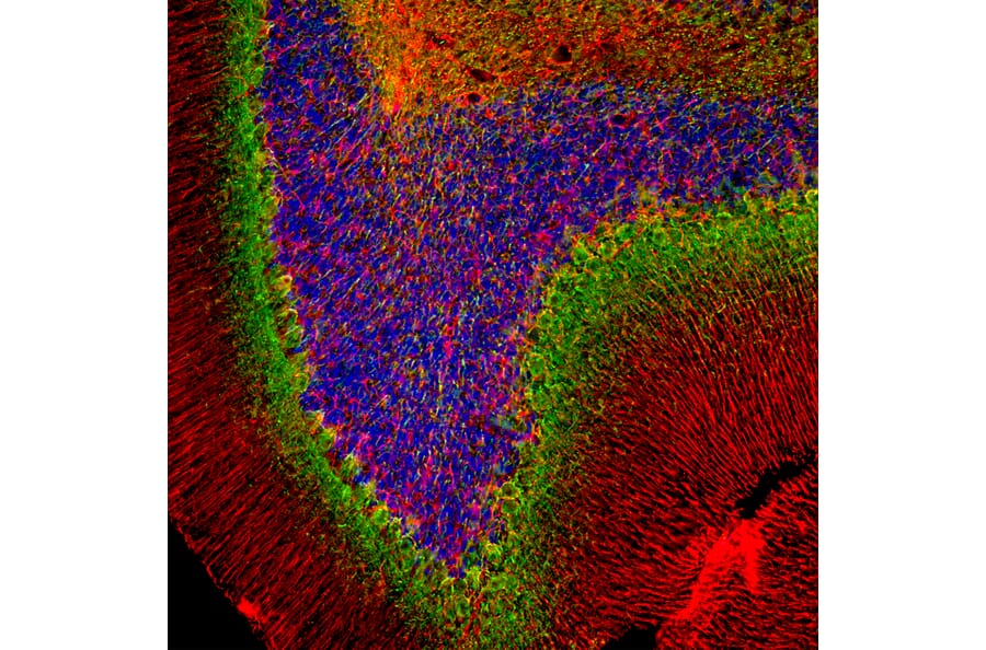
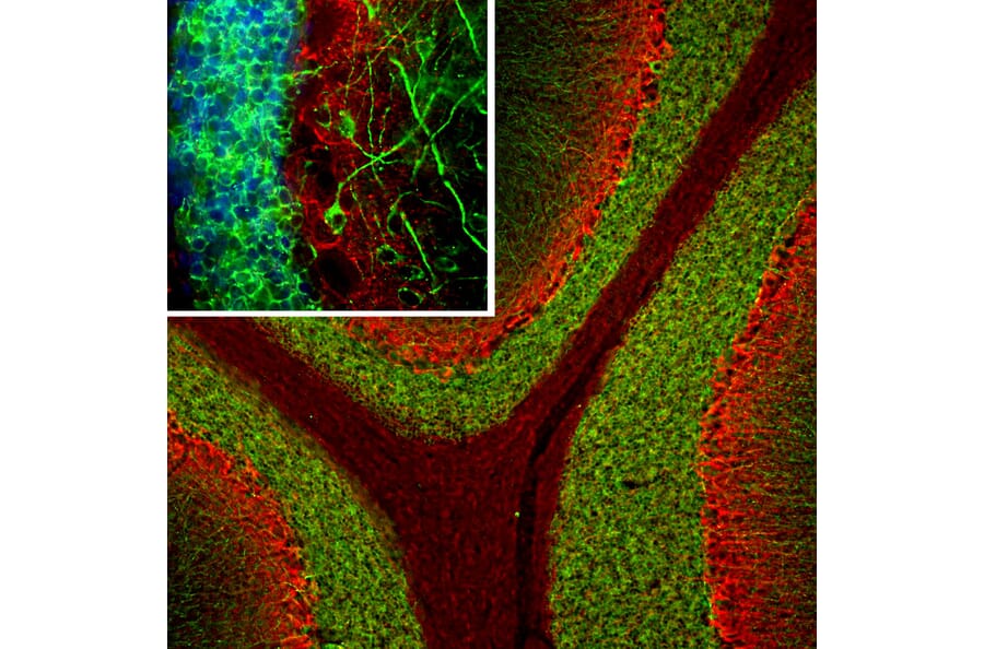
Immunofluorescent analysis of rat cerebellum section stained with Anti-Alpha-Internexin Antibody, at a dilution of 1:2,000, in green, and Anti-GFAP Antibody (A85307 | 1:5,000, in red. Blue is DAPI staining of nuclear DNA. Following transcardial perfusion with 4% paraformaldehyde, brain was post fixed for 24 hours, cut to 45 µM, and free-floating sections were stained with the above antibodies. The Anti-Alpha-Internexin Antibody selectively stains axons and dendrites of neuronal cells, in particular Purkinje cells and parallel fibers the axons of granule cells. The Anti-GFAP Antibody labels network of glial cells, such as astrocytes in the granule cell layer and white matter and Bergmann glia in the molecular layer.
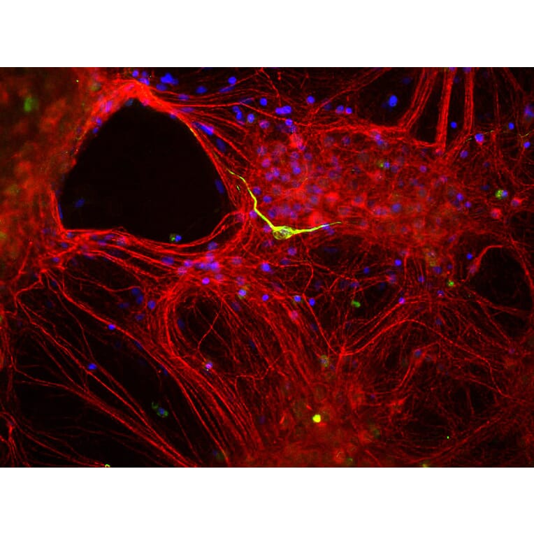
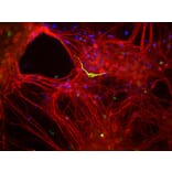
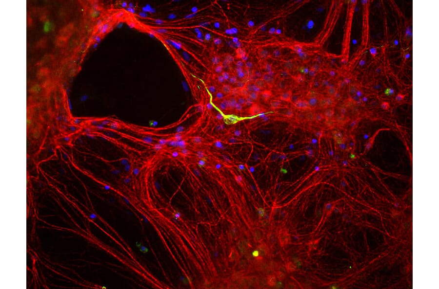
Mixed neuron-glial cultures stained with Anti-Alpha-Internexin Antibody (red) and Anti-Peripherin Antibody (A85435 | green). The Anti-Alpha-Internexin Antibody stains numerous axonal and dendritic profiles in these cultures, while Anti-Peripherin Antibody binds to only a subset of neurons.
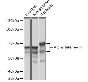
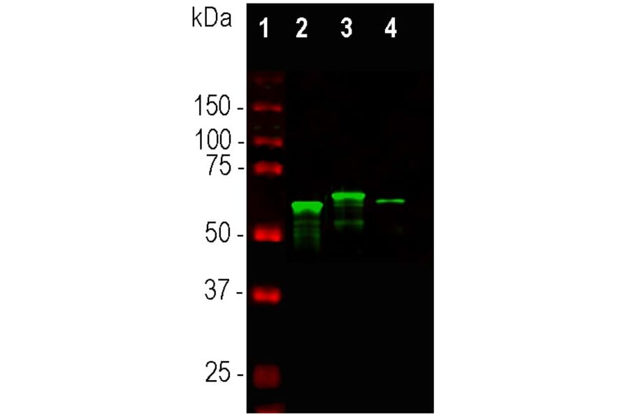
Western Blot - Anti-alpha Internexin Antibody (A85441)
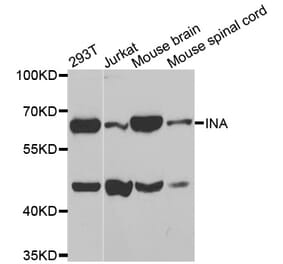
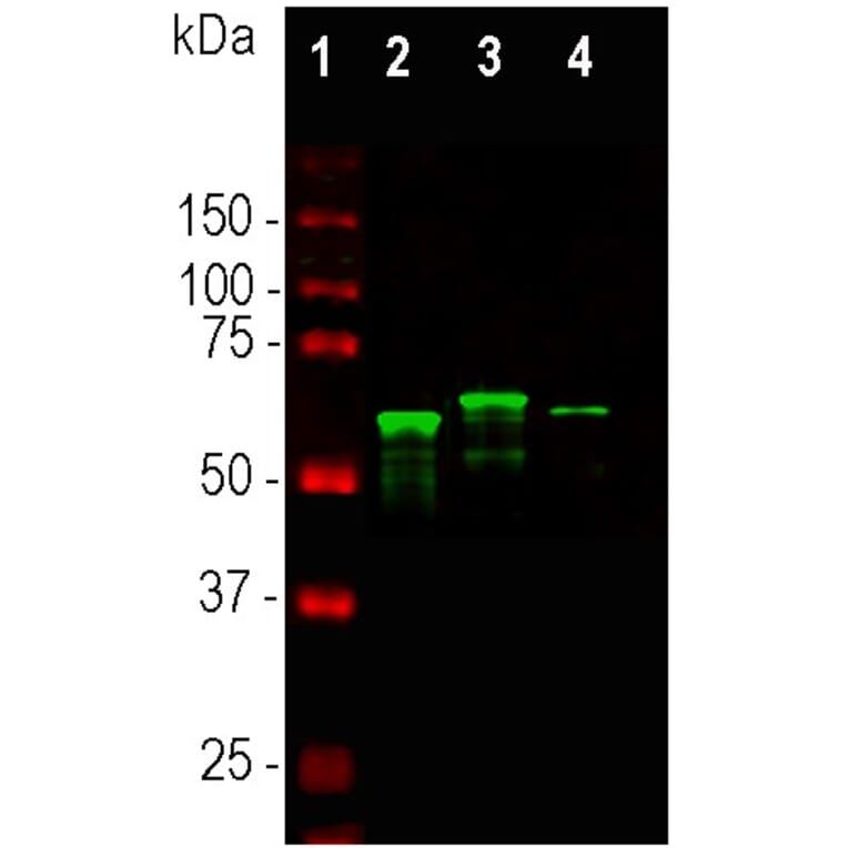
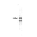
Western blot analysis of whole tissue lysates using Anti-Alpha-Internexin Antibody, at a dilution of 1:10,000, in green,: [Lane 1] protein standard (red), [Lane 2] mouse spinal cord, [Lane 3] rat spinal cord, [Lane 4] bovine spinal cord. Major bands in the 64-66 kDa range corresponds to a-internexin. The a-internexin protein from different species is known to vary slightly in SDS-PAGE molecular weight.
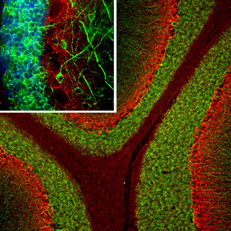
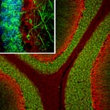
Immunofluorescent analysis of a rat cerebellum section stained with Anti-MAP2 Antibody [5H11] (A85296) at a dilution of 1:5,000 (green), and costained with Anti-alpha Internexin Antibody () at a dilution of 1:2,000 (red). Following transcardial perfusion of rat with 4% paraformaldehyde, brain was post fixed for 24 hours, cut to 45 µM, and free-floating sections were stained with above antibodies. The Anti-MAP2 Antibody [5H11] (A85296) antibody labels MAP2 protein in the perikarya and dendrites of the most neurons, particularly in granular cell and molecular layers of the cerebellum, while the a-internexin antibody selectively stains axons and dendrites of neuronal cells.
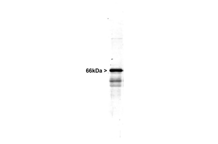
Western Blot - Anti-alpha Internexin Antibody (A85441)
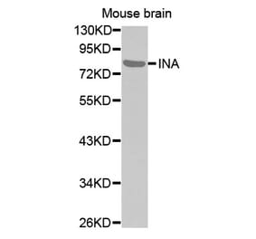
Western blot of whole rat spinal cord homogenate stained with Anti-Alpha-Internexin Antibody, at a dilution of 1:20,000). A prominent band running at ~66 kDa is apparent, as well as smaller lower bands which are apparently degradation products. A minor band at ~150 kDa is also seen, apparently resulting from dimerization of alpha-internexin.
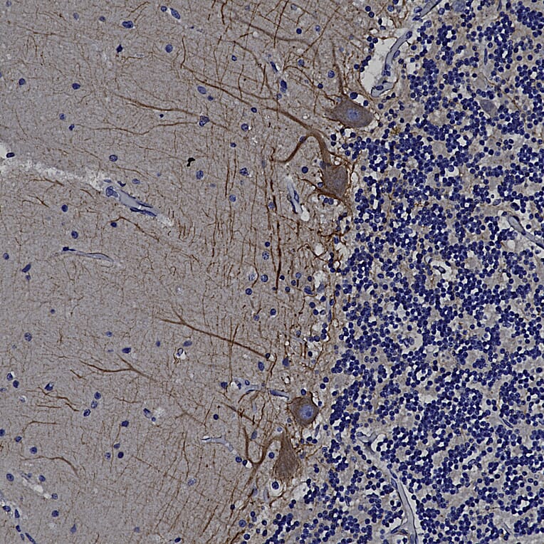
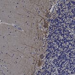
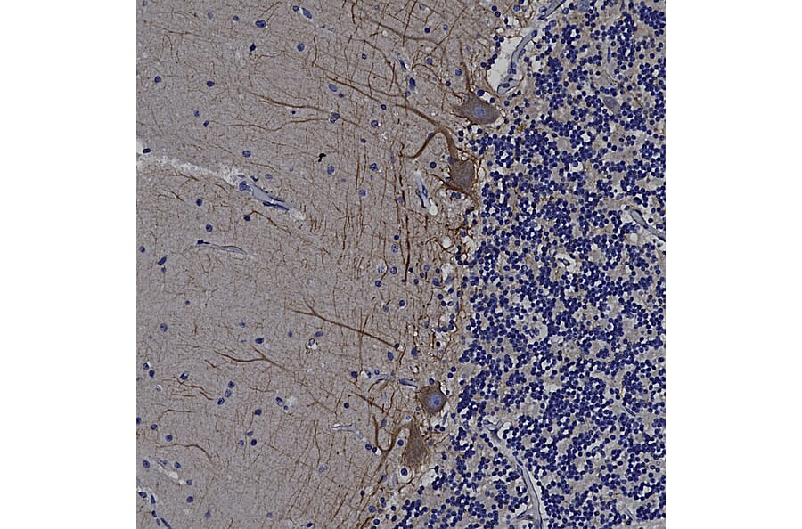
Immunohistochemistry analysis of a formalin fixed paraffin embedded human cerebellum section with Anti-alpha Internexin Antibody (A85441), detected with DAB (brown) using the Vector Labs ImmPRESS method and reagents with citra buffer retrieval. Counterstained with Hematoxylin (blue). The Anti-alpha Internexin Antibody (A85441) labels axons and dendrites of neuronal cells, outlining the Purkinje cells and parallel fibers in this image. Note: this antibody performs well in testing with both 4% PFA and standard NBF fixed rat, mouse, and human tissues.


![Immunohistochemistry - Anti-alpha Internexin Antibody [2E3] (A85448) - Antibodies.com](https://cdn.antibodies.com/image/catalog/85/A85448_1.jpg?profile=product_alternative)
![Immunofluorescence - Anti-alpha Internexin Antibody [1D2] (A85447) - Antibodies.com](https://cdn.antibodies.com/image/catalog/85/A85447_1.jpg?profile=product_alternative)