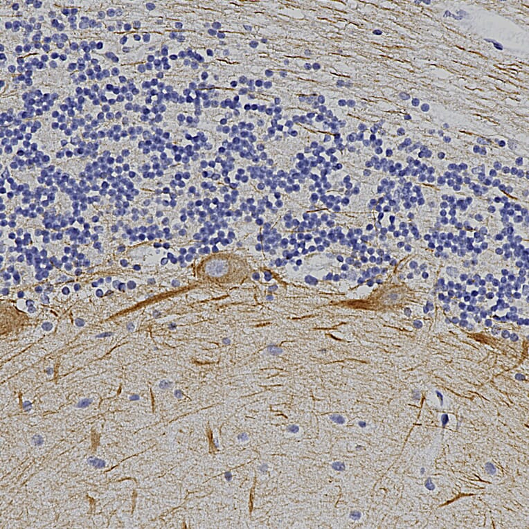
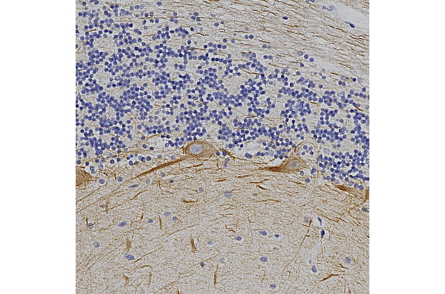
Immunohistochemistry analysis of a formalin fixed paraffin embedded human cerebellum section with Anti-alpha Internexin Antibody (A85446) at a dilution of 1:10,000 detected in DAB (brown) following the ABC method. Counterstained with Hematoxylin (blue). The a-internexin antibody selectively stains axons and dendrites of neuronal cells, in particular Purkinje cells, parallel fibers and the axons of granule cells.
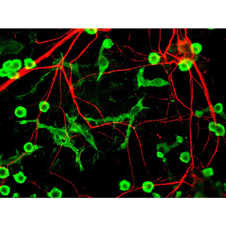
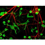
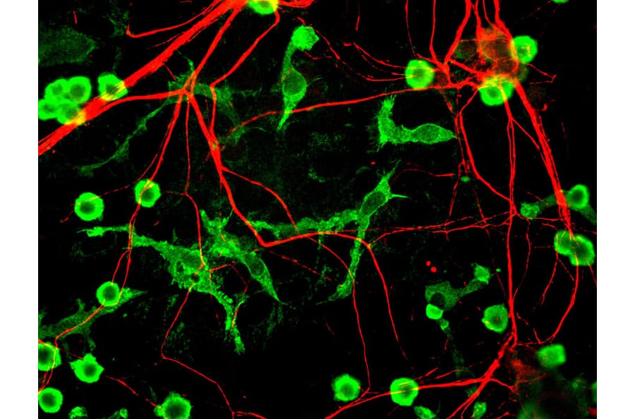
Mixed neuron glia cultures stained with Anti-Alpha-Internexin Antibody (red) and Anti-Coronin 1a Antibody (A85431 | green) - an excellent marker of microglia and lymphocytes. The Anti-Alpha-Internexin Antibody is an excellent marker of neuronal processes in these cultures.
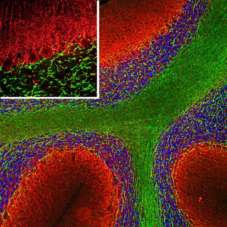
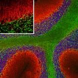
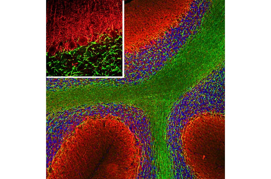
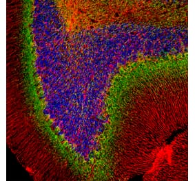
Immunofluorescent analysis of a rat cerebellum section stained with Anti-alpha Internexin Antibody (A85446), at a dilution of 1:5,000, in red, and co-stained with Anti-MBP Antibody (A85322), at a dilution of 1:5,000, in green. The nuclear DNA is visualised in blue using Hoechst staining. Following transcardial perfusion of the rat with 4% paraformaldehyde, the brain was post-fixed for 24 hours, cut to 45 µm, and free-floating sections were stained with the above antibodies. The Anti-alpha Internexin Antibody (A85446) selectively stains axons and dendrites of neuronal cells, in particular Purkinje cells, parallel fibers and the axons of granule cells, while the Anti-MBP Antibody (A85322) stains myelin sheathes around axons.
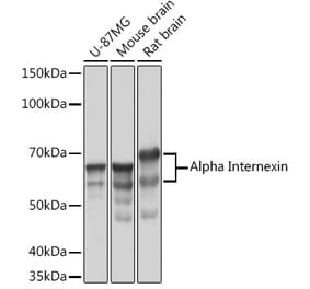
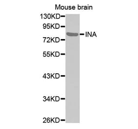
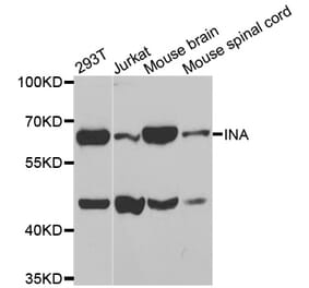
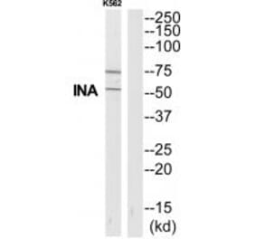
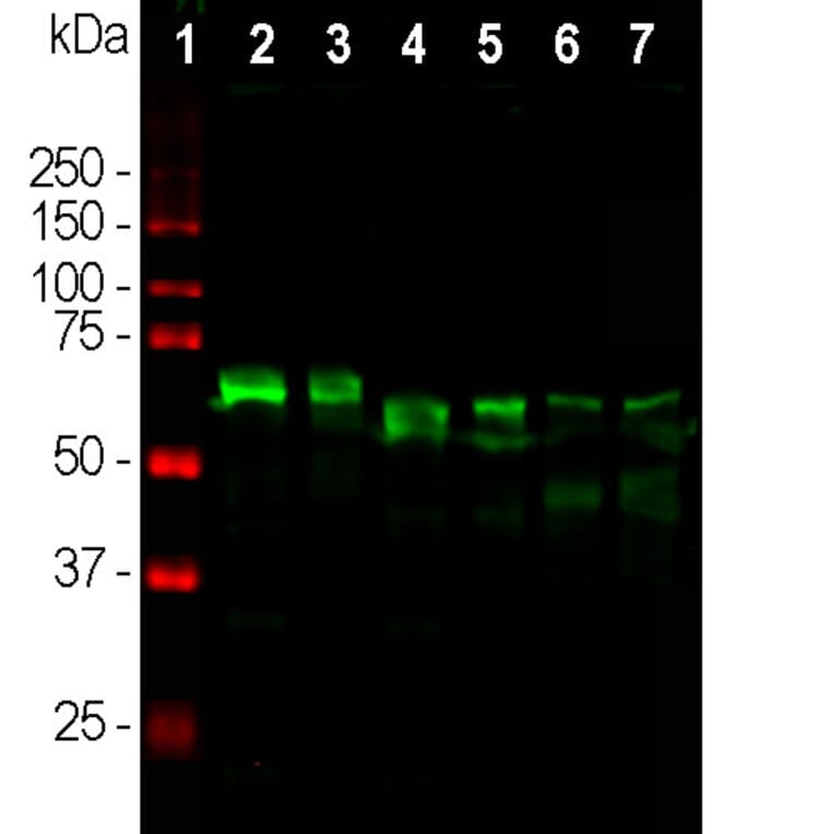
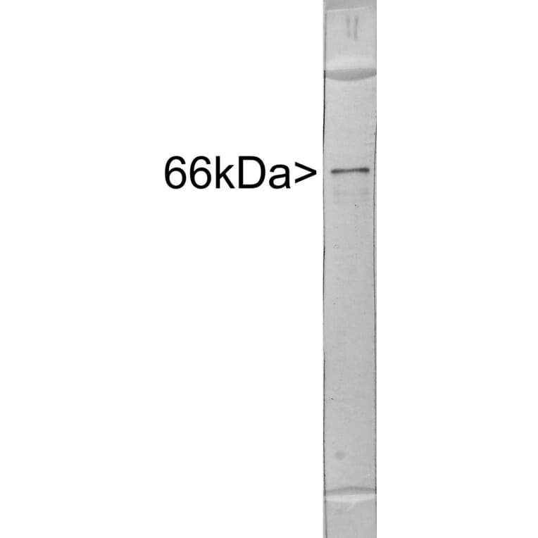
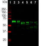
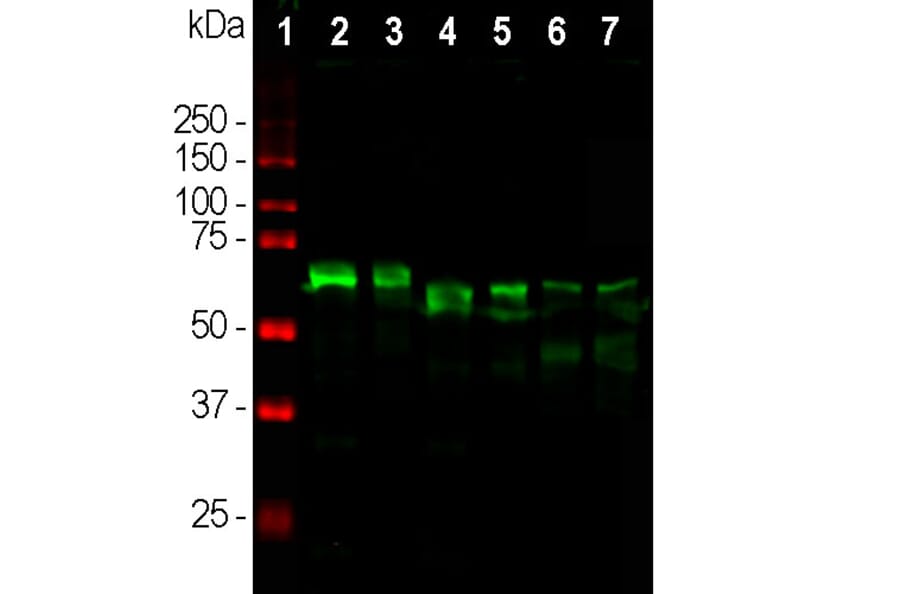
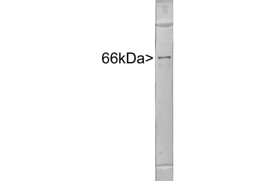
Western Blot - Anti-alpha Internexin Antibody (A85446)
Western blot analysis of different tissue lysates using Anti-alpha Internexin Antibody (A85446), at a dilution of 1:10,000, in green. The lanes contain samples of: [Lane 1] Protein standards, in red, [Lane 2] rat brain, [Lane 3] rat spinal cord, [Lane 4] mouse brain, [Lane 5] mouse spinal cord, [Lane 6] cow spinal cord and [Lane 7] pig spinal cord. The Anti-alpha Internexin Antibody (A85446) reveals the alpha Internexin protein with apparent molecular weight of 64-66 kDa, with some variability among different species.


![Immunohistochemistry - Anti-alpha Internexin Antibody [2E3] (A85448) - Antibodies.com](https://cdn.antibodies.com/image/catalog/85/A85448_1.jpg?profile=product_alternative)
![Immunofluorescence - Anti-alpha Internexin Antibody [1D2] (A85447) - Antibodies.com](https://cdn.antibodies.com/image/catalog/85/A85447_1.jpg?profile=product_alternative)