This antibody recognises bromodeoxyuridine (known as BrdU or BrdUrd). BrdU is a synthetic thymidine analog, which when added to cell culture media can be incorporated into the DNA of replicating cells as a substitute for thymidine. This characteristic combined with the ease of detecting incorporated BrdU with the help of anti-BrdU antibodies has resulted in establishing BrdU as a key tool for the visualization of cells in the S-Phase of the cell cycle. BrdU is also commonly used in vivo for monitoring cell proliferation in model systems; BrdU can be administered via intraperitoneal injection, subcutaneous osmotic pump implants or in drinking water.
Applications
IHC-P, IF, Flow Cytometry, ICC
Dilutions
FC 1: 1:50 - 1:100, ICC: 1:500, IF 2: 1:100 - 1:500, IHC-P: 1:100 - 1:500
Reactivity
Species Independent
Immunogen
Bromodeoxyuridine conjugated to Keyhole Limpet Haemocyanin.
Host
Rabbit
Clonality
Polyclonal
Isotype
IgG
Conjugate
Unconjugated
Concentration
1 mg/ml
Product Form
Liquid
Formulation
Supplied in Phosphate Buffered Saline with <0.1% Sodium Azide.
Storage
Store undiluted at -20°C only. Storage in frost free freezers is not recommended. Avoid freeze-thaw cycles. Should this product contain a precipitate we recommend microcentrifugation before use.
General Notes
Rabbit anti BrdU antibody recognizes bromodeoxyuridine (known as BrdU or BrdUrd). BrdU is a synthetic thymidine analog, which when added to cell culture media can be incorporated into the DNA of replicating cells as a substitute for thymidine. This characteristic combined with the ease of detecting incorporated BrdU with the help of anti-BrdU antibodies has resulted in establishing BrdU as a key tool for the visualization of cells in the S-Phase of the cell cycle. BrdU is also commonly used in vivo for monitoring cell proliferation in model systems; BrdU can be administered via intraperitoneal injection, subcutaneous osmotic pump implants (Tesfaiqzi et al. 2004) or in drinking water (Moser et al. 2004).
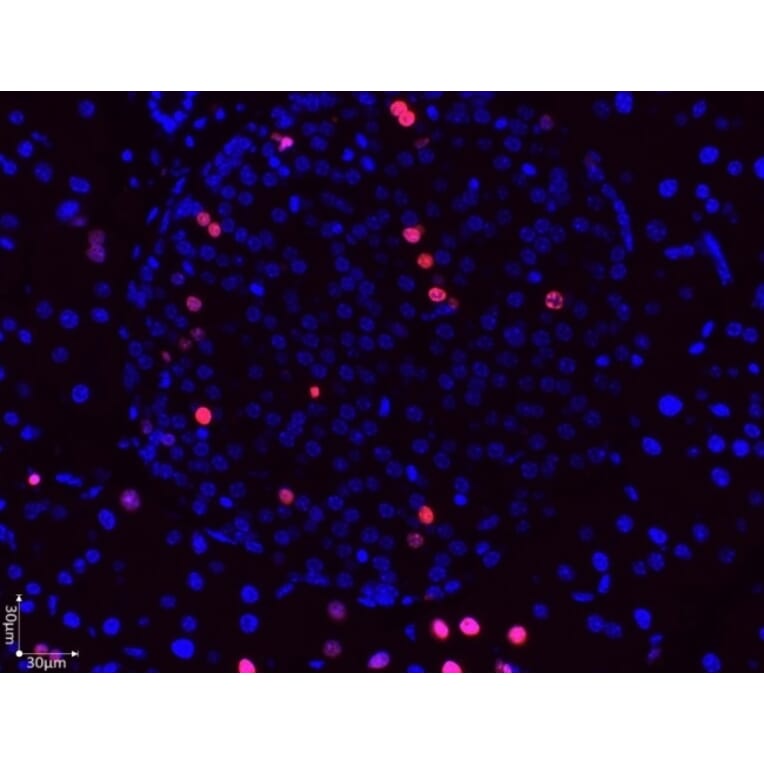
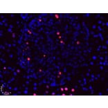
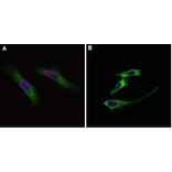
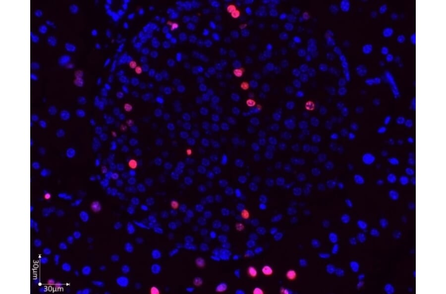
Immunofluorescence microscopy image of paraformaldehyde-fixed, paraffin-embedded guinea pig pancreas tissue stained with 2.7 µ/ml Anti-BrdU Antibody (A283414). Donkey anti-rabbit antibody was used as the secondary antibody (red). DAPI was used as nuclear counterstain (blue) and imaged with a 40x objective.
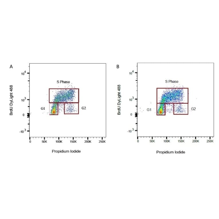
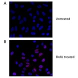
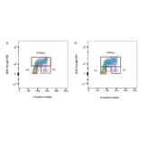
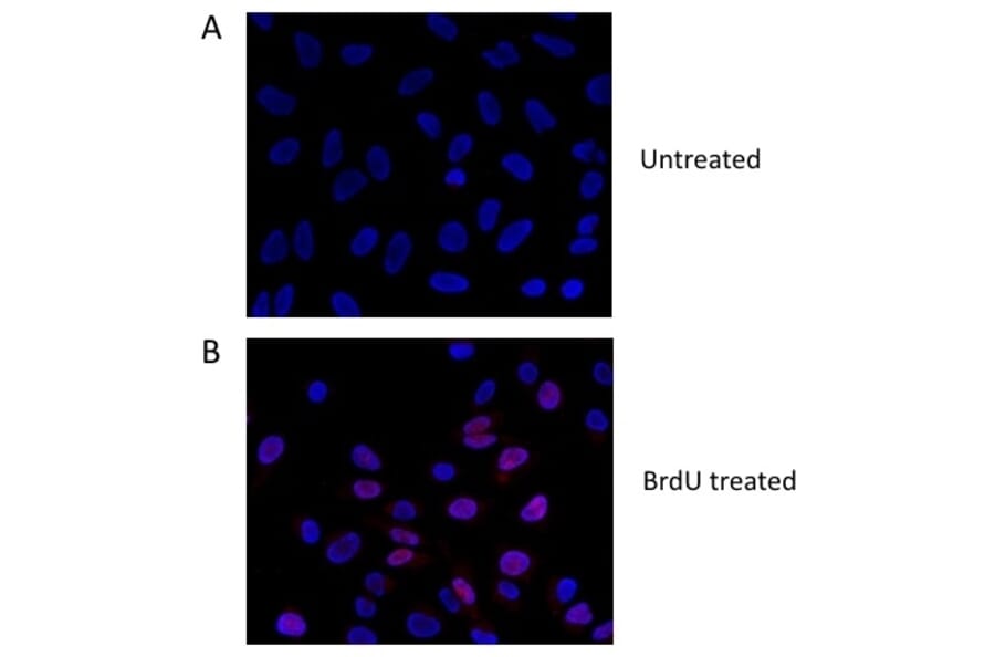
BrdU-labeled human lymphoma cells were stained with Anti-BrdU Antibody (A283414) at a 1/50 dilution (A) and a 1/100 dilution (B). Sheep anti Rabbit IgG DyLight 488 conjugated antibody was used as the secondary detection reagent at a 1/50 dilution. Propidium Iodide was used to stain total DNA.
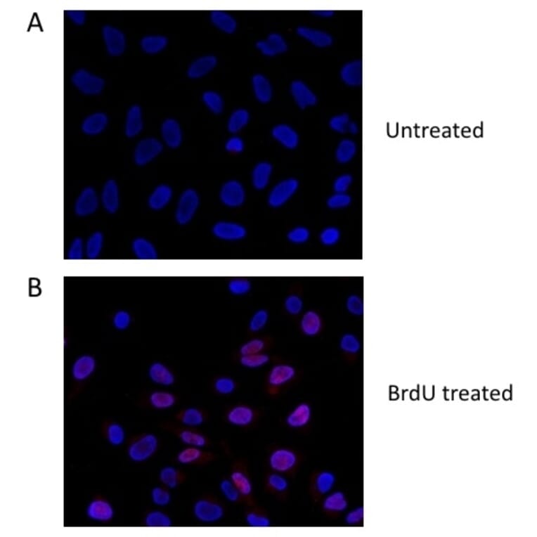
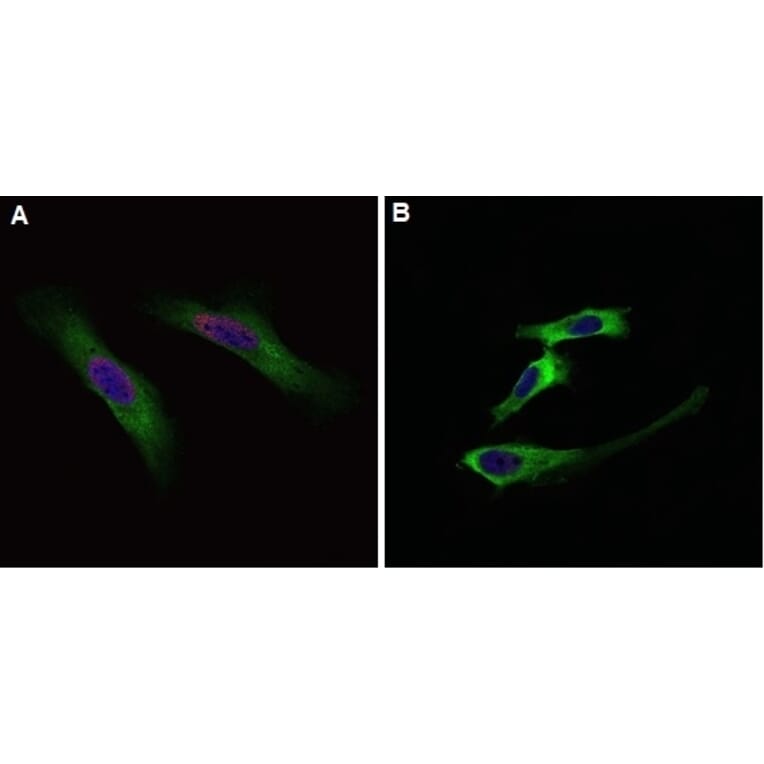
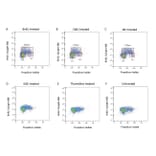
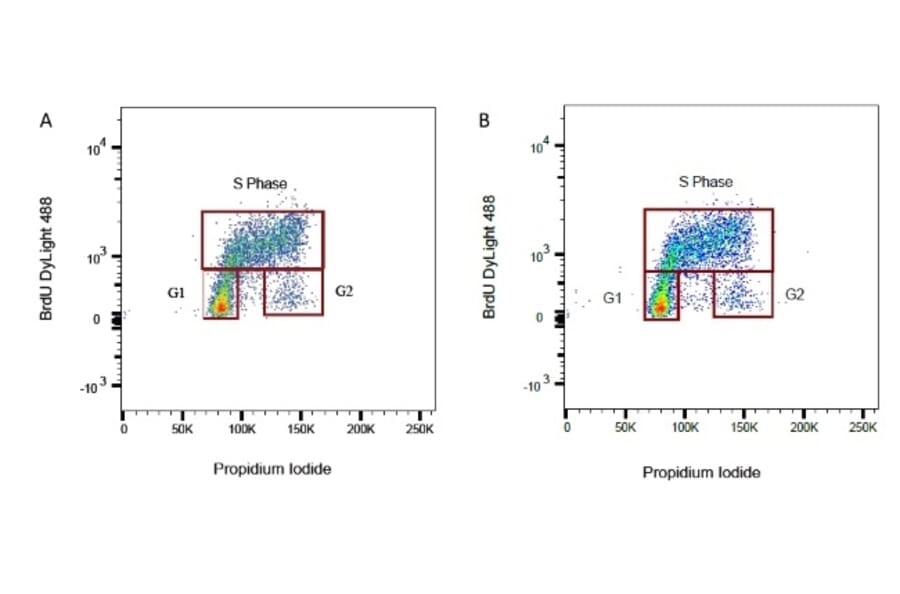
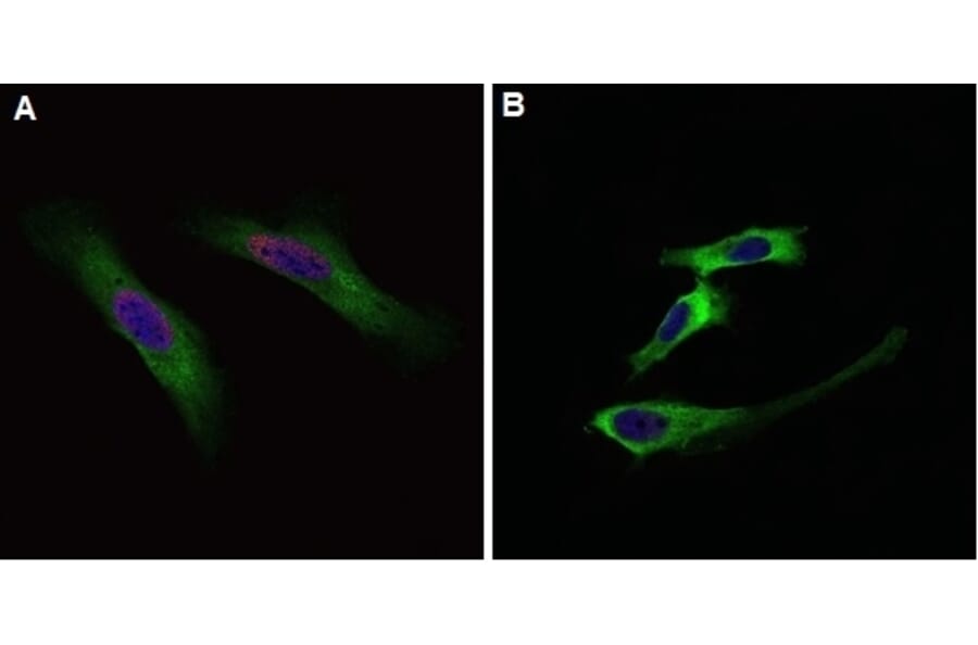
Flow cytometric analysis of HeLa cells treated with BrdU (A) or DMSO (B) and stained with Anti-BrdU Antibody (A283414) at a 1/100 dilution. Sheep anti Rabbit IgG DyLight 549 conjugated antibody (red) at a 1/500 dilution was used as the secondary antibody. Cytoplasm was stained with Mouse anti Human GAPDH antibody at a 1/500 dilution. As the secondary antibody, Goat anti Mouse IgG (H/L) DyLight 488 conjugated antibody (green) was used at a 1/500 dilution. PureBlu DAPI was used as nuclear counterstain (blue).
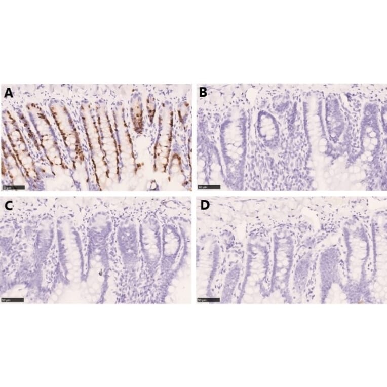
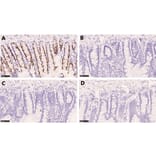
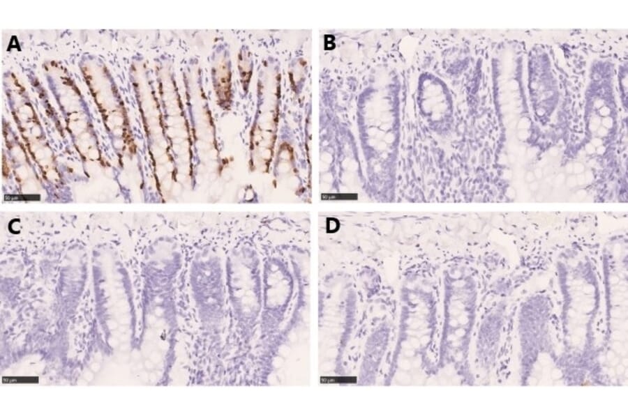
Immunohistochemistry analysis showing staining of BrdU with Anti-BrdU Antibody (A283414) in the nucleus of paraffin-embedded Rat Jejunum treated with 100 mg/kg BrdU in PBS (A) compared to Rat Jejunum treated with PBS only (C). A negative control without primary antibody was also included for both BrdU treated (B) and untreated tissue (D). Detection was performed using an HRP conjugated secondary antibody followed by colorimetric detection using DAB. Tissues were counterstained with hematoxylin.


![Flow Cytometry - Anti-BrdU Antibody [MoBu-1] (A85813)](https://cdn.antibodies.com/image/catalog/85/A85813_1.jpg?profile=product_alternative)
![Flow Cytometry - Anti-BrdU Antibody [Bu20a] (A86223)](https://cdn.antibodies.com/image/catalog/86/A86223_1.jpg?profile=product_alternative)
![Flow Cytometry - Anti-BrdU Antibody [IIB5] (A115514) - Antibodies.com](https://cdn.antibodies.com/image/catalog/115/A115514_1.jpg?profile=product_alternative)
![Flow Cytometry - Anti-BrdU Antibody [Bu20a] (A279885) - Antibodies.com](https://cdn.antibodies.com/image/catalog/279/A279885_1.jpg?profile=product_alternative)
![Immunohistochemistry - Anti-BrdU Antibody [BRD.3] - BSA and Azide free (A250986) - Antibodies.com](https://cdn.antibodies.com/image/catalog/254/A254166_1.jpg?profile=product_alternative)
![SDS-PAGE - Anti-BrdU Antibody [MoBu-1] (A254186) - Antibodies.com](https://cdn.antibodies.com/image/catalog/250/A250978_1.jpg?profile=product_alternative)
![SDS-PAGE - Anti-BrdU Antibody [MoBu-1] - BSA and Azide free (A250978) - Antibodies.com](https://cdn.antibodies.com/image/catalog/254/A254158_1.jpg?profile=product_alternative)
![Immunohistochemistry - Anti-BrdU Antibody [BRD.3] (A254173) - Antibodies.com](https://cdn.antibodies.com/image/catalog/250/A250986_1.jpg?profile=product_alternative)
![Immunohistochemistry - Anti-BrdU Antibody [BRD469] (A254176) - Antibodies.com](https://cdn.antibodies.com/image/catalog/250/A250878_1.jpg?profile=product_alternative)
![Immunohistochemistry - Anti-BrdU Antibody [BRD469] - BSA and Azide free (A250878) - Antibodies.com](https://cdn.antibodies.com/image/catalog/254/A254058_1.jpg?profile=product_alternative)
![Immunohistochemistry - Anti-BrdU Antibody [85-2C8] (A253608) - Antibodies.com](https://cdn.antibodies.com/image/catalog/250/A250994_1.jpg?profile=product_alternative)
![Immunohistochemistry - Anti-BrdU Antibody [85-2C8] - BSA and Azide free (A250994) - Antibodies.com](https://cdn.antibodies.com/image/catalog/254/A254173_1.jpg?profile=product_alternative)
![Immunohistochemistry - Anti-BrdU Antibody [SPM166] (A251240) - Antibodies.com](https://cdn.antibodies.com/image/catalog/250/A250906_1.jpg?profile=product_alternative)
![Immunohistochemistry - Anti-BrdU Antibody [SPM166] - BSA and Azide free (A250906) - Antibodies.com](https://cdn.antibodies.com/image/catalog/254/A254086_1.jpg?profile=product_alternative)