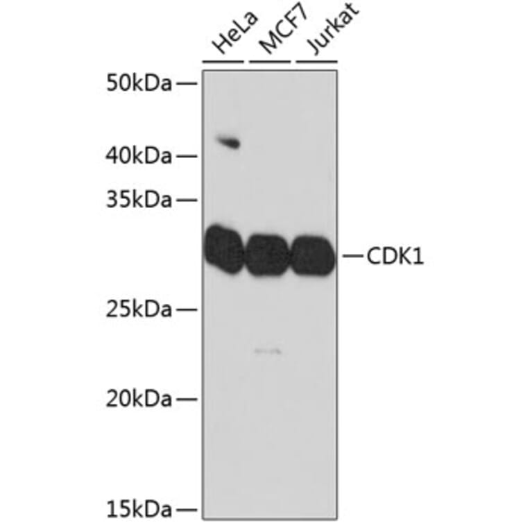
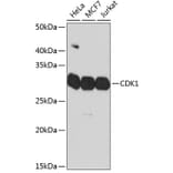
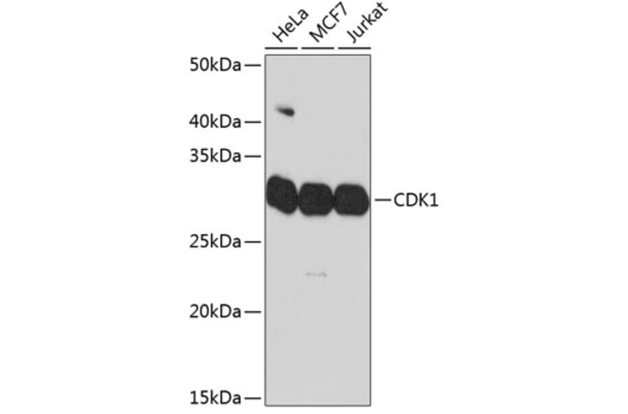
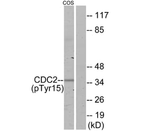
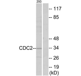
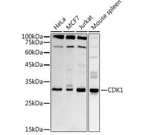
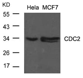
Western Blot - Anti-CDK1 Antibody [ARC50607] (A305462)
Western blot analysis of extracts of various cell lines, using Anti-CDK1 Antibody (A305462) at 1:1,000 dilution. The secondary antibody was Goat Anti-Rabbit IgG H&L Antibody (HRP) at 1:10,000 dilution. Lysates/proteins were present at 25µg per lane. The blocking buffer used was 3% non-fat dry milk in TBST. Detection was with a ECL Basic Kit. Exposure time: 90s.
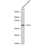
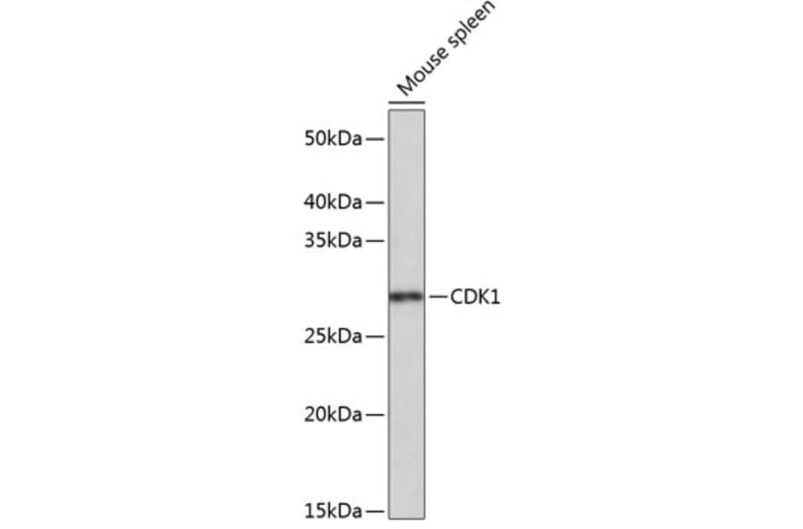
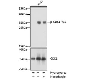
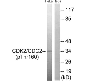
Western Blot - Anti-CDK1 Antibody [ARC50607] (A305462)
Western blot analysis of extracts of Mouse spleen, using Anti-CDK1 Antibody (A305462) at 1:1,000 dilution. The secondary antibody was Goat Anti-Rabbit IgG H&L Antibody (HRP) at 1:10,000 dilution. Lysates/proteins were present at 25µg per lane. The blocking buffer used was 3% non-fat dry milk in TBST. Detection was with a ECL Basic Kit. Exposure time: 3min.
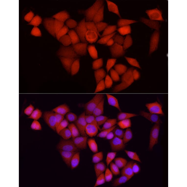
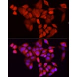
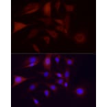
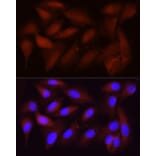
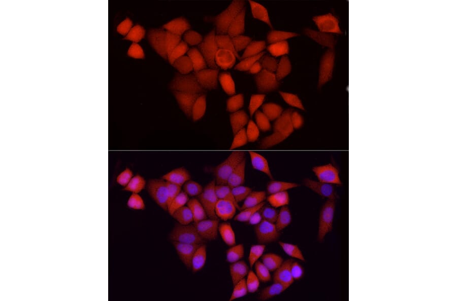
Immunofluorescence analysis of HeLa using Anti-CDK1 Antibody (A305462) at a dilution of 1:100 (40x lens). DAPI was used to stain the cell nuclei (blue).
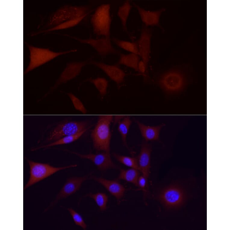
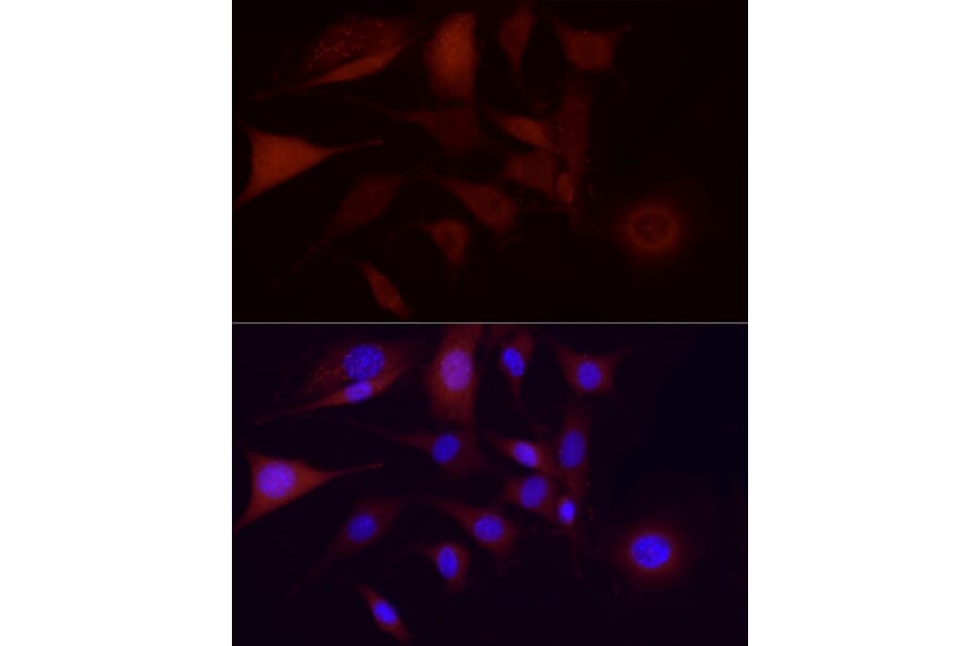
Immunofluorescence analysis of NIH/3T3 using Anti-CDK1 Antibody (A305462) at a dilution of 1:100 (40x lens). DAPI was used to stain the cell nuclei (blue).
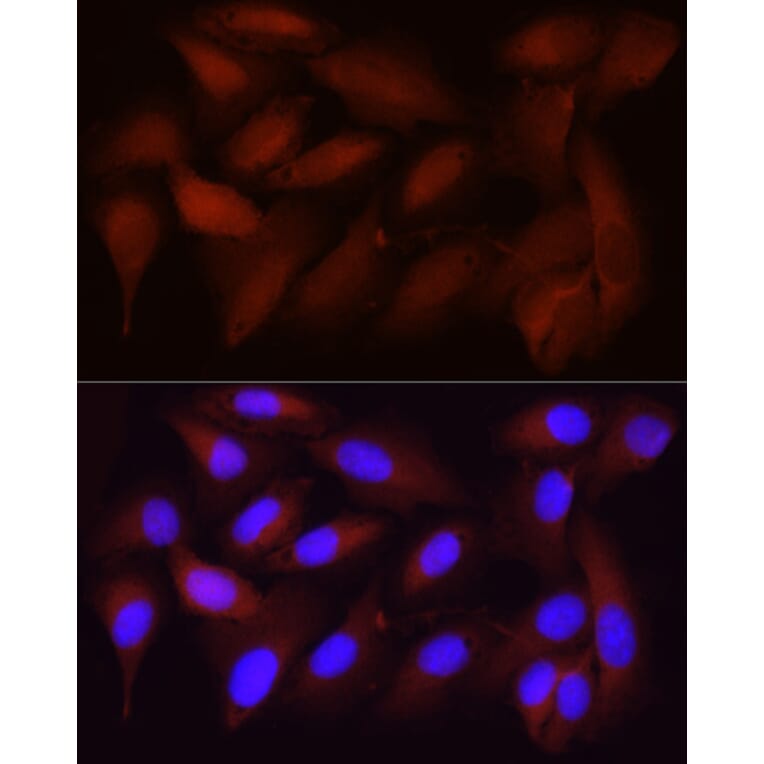
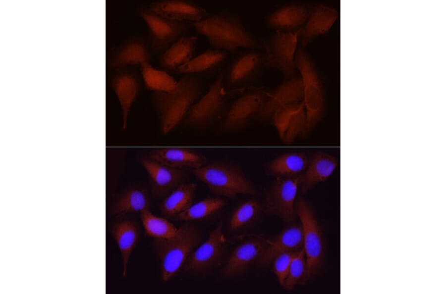
Immunofluorescence analysis of U2OS using Anti-CDK1 Antibody (A305462) at a dilution of 1:100 (40x lens). DAPI was used to stain the cell nuclei (blue).
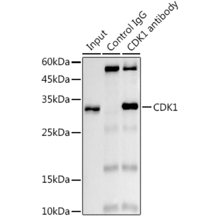
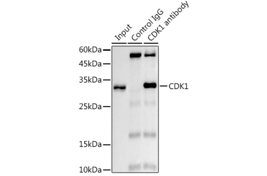
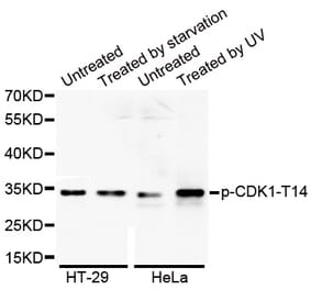
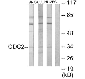
Western Blot - Anti-CDK1 Antibody [ARC50607] (A305462)
Immunoprecipitation analysis of 300µg extracts of 293T cells using 3µg of Anti-CDK1 Antibody (A305462). This Western blot was performed on the immunoprecipitate using Anti-CDK1 Antibody (A305462) at a dilution of1:1000.


![IHC - Anti-CDK1 Antibody [POH-1] (A86650)](https://cdn.antibodies.com/image/catalog/86/A86650_1.jpg?profile=product_alternative)
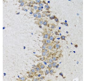
![Immunohistochemistry - Anti-CDK1 Antibody [A17.1.1] (A253992) - Antibodies.com](https://cdn.antibodies.com/image/catalog/250/A250808_1.jpg?profile=product_alternative)
![Immunohistochemistry - Anti-CDK1 Antibody [A17.1.1] - BSA and Azide free (A250808) - Antibodies.com](https://cdn.antibodies.com/image/catalog/253/A253988_1.jpg?profile=product_alternative)
![Immunohistochemistry - Anti-CDK1 Antibody [POH-1] (A253987) - Antibodies.com](https://cdn.antibodies.com/image/catalog/250/A250806_1.jpg?profile=product_alternative)