Unconjugated
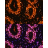
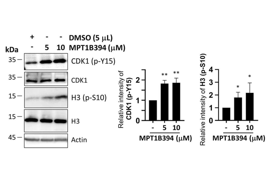
Glioblastoma (GBM) is a highly aggressive and therapeutically challenging brain tumor characterized by poor prognosis, rapid recurrence, and resistance to standard treatments such as temozolomide (TMZ). Novel therapeutic strategies that target core survival and proliferative pathways in GBM are urgently needed. In this study, we synthesized and screened a series of dual CDC25-HDAC inhibitors for anti-GBM activity. Among them, MPT1B394 emerged as a potent candidate, exhibiting strong cytotoxicity in both parental and TMZ-resistant GBM cell lines, along with favorable in vivo safety and survival outcomes in an orthotopic GBM mouse model. Transcriptomic profiling via RNA sequencing revealed that MPT1B394 treatment significantly downregulated the cell cycle checkpoint pathway. More than twenty mitosis-associated genes were suppressed in both parental and resistant A172 cells. Integration with GBM patient datasets confirmed that elevated expression of these genes, including BIRC5, CDCA8, PLK1, CCNB1, CCNA2, MCM5, and CENPA, correlates with poor patient survival. Blood-brain barrier (BBB) penetration predictions supported MPT1B394's ability to access intracranial tumors, reinforcing its potential as a central nervous system-active agent. Protein-level validation through immunofluorescence and analysis of public proteomic datasets demonstrated markedly higher expression of chromosomal passenger complex (CPC) components (BIRC5 and Aurora B) in GBM specimens, which were notably reduced following MPT1B394 treatment. Disruption of CPC function led to reduced mitotic activity and increased mitotic defects in treated cultures. Collectively, these findings indicate that dual inhibition of CDC25 and HDAC by MPT1B394 disrupts CPC-mediated mitosis and aberrant cell cycle progression, representing a promising therapeutic avenue for GBM treatment.


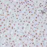

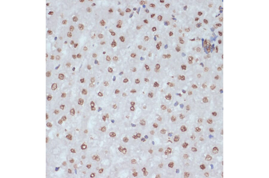
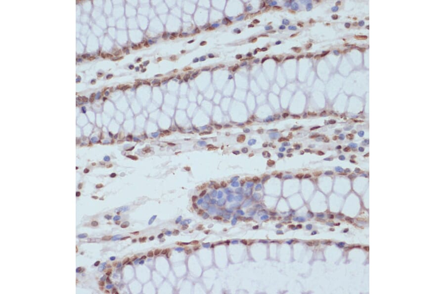

![IHC - Anti-CDK1 Antibody [POH-1] (A86650)](https://cdn.antibodies.com/image/catalog/86/A86650_1.jpg?profile=product_alternative)
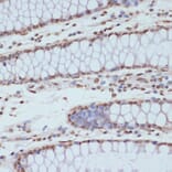
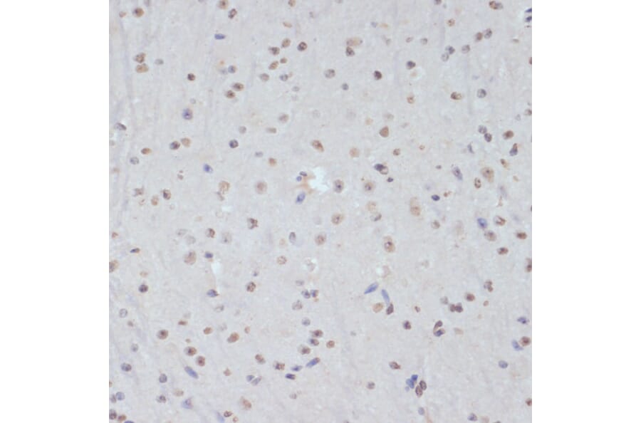
![Immunohistochemistry - Anti-CDK1 Antibody [A17.1.1] (A253992) - Antibodies.com](https://cdn.antibodies.com/image/catalog/250/A250808_1.jpg?profile=product_alternative)
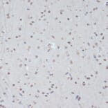
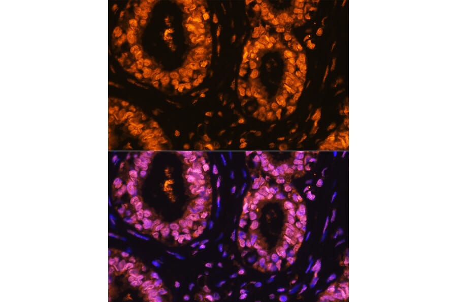
![Immunohistochemistry - Anti-CDK1 Antibody [A17.1.1] - BSA and Azide free (A250808) - Antibodies.com](https://cdn.antibodies.com/image/catalog/253/A253988_1.jpg?profile=product_alternative)